Bacterial microscopic image segmentation method based on deep learning network

A deep learning network and microscopic image technology, applied in the field of bacterial microscope image segmentation, can solve the problems of tediousness and poor segmentation effect, and achieve the effect of reducing the amount of calculation, excellent image segmentation effect, and increasing size

Pending Publication Date: 2021-06-11
至微生物智能科技(厦门)有限公司
View PDF1 Cites 3 Cited by
  • Summary
  • Abstract
  • Description
  • Claims
  • Application Information

AI Technical Summary

Problems solved by technology

[0018] It does a lot of work for image preprocessing, which is relatively cumbersome. If these contrast enhancements are not performed, the segmentation effect will be poor.

Method used


Image

  • Bacterial microscopic image segmentation method based on deep learning network
  • Bacterial microscopic image segmentation method based on deep learning network
  • Bacterial microscopic image segmentation method based on deep learning network

Examples


Embodiment Construction

[0048] The present invention will be further described below through specific embodiments.

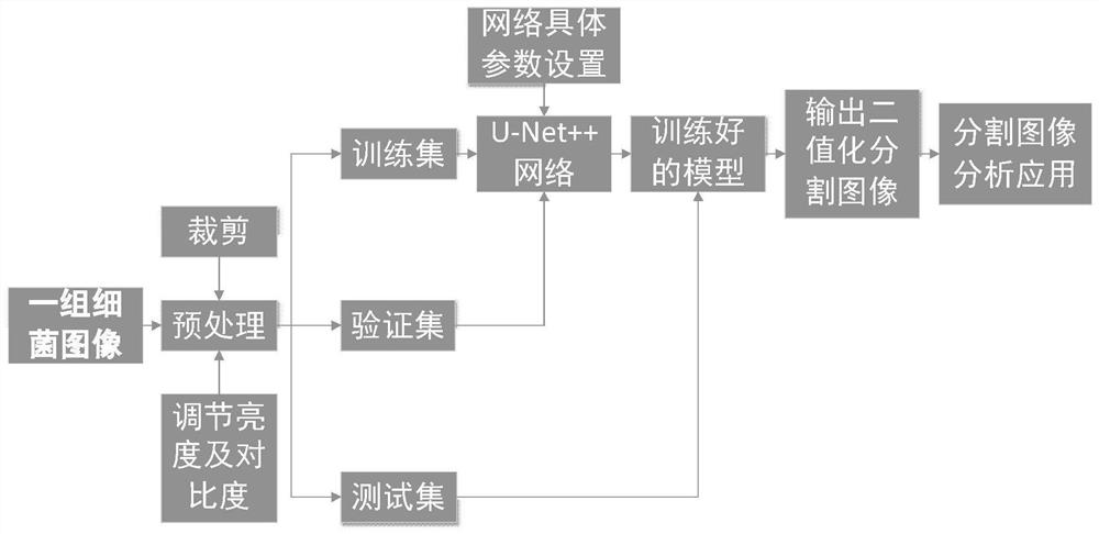
[0049] see figure 1 , a bacterial microscopic image segmentation method based on a deep learning network, comprising the following steps:

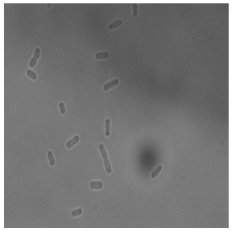
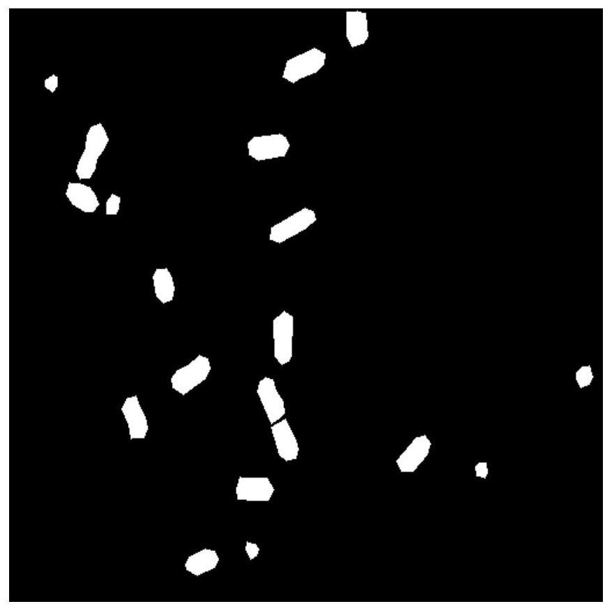
[0050] 1) Cultivate bacteria, and take a set of pictures of bacterial growth under a microscope at fixed time intervals, perform image preprocessing, and construct a training set, a verification set, and a test set that do not overlap with each other, and the training set includes the original image and the corresponding The labeled images, validation set and test set respectively contain only original images.

[0051] Because the pixel values ​​of the images taken by bacteria under the microscope are generally large, and the quality of the images varies, so it is necessary to perform simple cropping, brightness and contrast enhancement preprocessing on the images before deep learning. Specifically include the following:

[0052] 1.1) Cultivate ...


PUM


Abstract

A bacteria microscopic image segmentation method based on a deep learning network comprises the following steps: 1) culturing bacteria, shooting a group of bacteria growth pictures under a microscope according to a fixed time interval, carrying out image preprocessing, constructing a training set, a verification set and a test set which are not intersected with one another, wherein the training set comprises original images and corresponding label images, and the verification set and the test set only comprise the original images respectively; (2) building a U-Net + + model, wherein the U-Net + + model is provided with an encoder module and a decoder module, the encoder module carries out feature extraction, the decoder module carries out feature reduction decoding to obtain the size of an original image; inputting the training set into the U-Net + + model for training, then inputting the verification set into the trained U-Net + + model for verification, and obtaining the trained U-Net + + model; and 3) inputting the test set into the trained U-Net + + model, and outputting a binary segmentation image. According to the method, the bacterial microscopic image can be automatically segmented quickly and accurately, too many complex image preprocessing links in the early stage are omitted, and time is saved.

Description

technical field [0001] The invention relates to the field of bacterial microscope image segmentation, in particular to a bacterial microscope image segmentation method based on a deep learning network. Background technique [0002] In the research and processing of images, the information contained in the images is often not what we are interested in. We will automatically judge which image information we need according to our needs, and often this specific information contains what we want. The image part of the information corresponds to the special properties (edge, shape, color, etc.) in the image. In biological image processing, we often call it the foreground, and the corresponding other image parts are the background. In the biological field, cultured bacteria are imaged under a microscope. These images are often affected by factors such as changes in the focal plane of the microscope, magazines in the culture medium, and environmental brightness. These noise-containi...

Claims


Application Information

Patent Timeline
Patent Type & Authority Applications(China)
IPC IPC(8): G06K9/00G06K9/34G06K9/62G06N3/04G06N3/08
CPCG06N3/08G06V20/69G06V20/695G06V10/267G06N3/045G06F18/253G06F18/214
Inventor 不公告发明人
Owner 至微生物智能科技(厦门)有限公司
Why Eureka
Try Eureka