Supercharge Your Innovation With Domain-Expert AI Agents!

Photoacoustic imaging method and system for mammary gland

A photoacoustic imaging and photoacoustic image technology, applied in the field of medical imaging, can solve the problems of photoacoustic image vibration error, slow imaging speed, photoacoustic signal over time, etc., and achieve the effect of improving positioning accuracy and accurate and clear collection

Pending Publication Date: 2021-06-18
XIEHE HOSPITAL ATTACHED TO TONGJI MEDICAL COLLEGE HUAZHONG SCI & TECH UNIV
View PDF5 Cites 0 Cited by
  • Summary
  • Abstract
  • Description
  • Claims
  • Application Information

AI Technical Summary

Problems solved by technology

[0007] The object of the present invention is to provide a photoacoustic imaging method and system for mammary glands to solve the problem that the prior art photoacoustic imaging method needs more time to collect photoacoustic signals due to the limitation of mechanical rotation, and the imaging speed is slow. At the same time, mechanical vibration will introduce vibration errors to photoacoustic images, which will bring instability to photoacoustic imaging.

Method used


Image

  • Photoacoustic imaging method and system for mammary gland
  • Photoacoustic imaging method and system for mammary gland
  • Photoacoustic imaging method and system for mammary gland

Examples


Embodiment 1

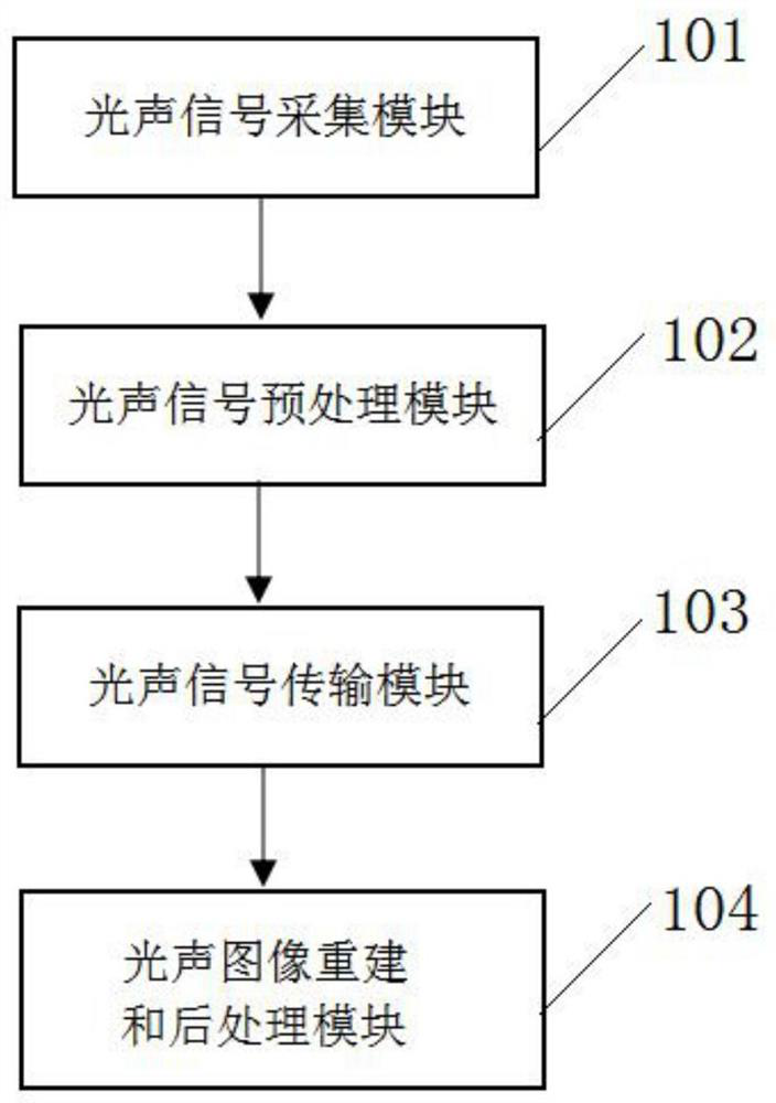
[0049] Such as figure 1 As shown, a photoacoustic imaging system for mammary glands, including: sequentially connected photoacoustic signal acquisition module, photoacoustic signal preprocessing module, photoacoustic signal transmission module, photoacoustic signal reconstruction and processing module, wherein:

[0050] A photoacoustic signal collection module 101, configured to collect photoacoustic signals generated after the laser irradiates breast tissue;

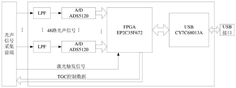
[0051] A photoacoustic signal preprocessing module 102, configured to preprocess the photoacoustic signal and convert it into a digital signal;

[0052] A photoacoustic signal transmission module 103, configured to transmit the preprocessed digital signal to the photoacoustic signal reconstruction and processing module;

[0053]The photoacoustic signal reconstruction and post-processing module 104 is used to reconstruct the optical coefficient absorption distribution of the breast tissue, and perform medical imaging on...

Embodiment 2

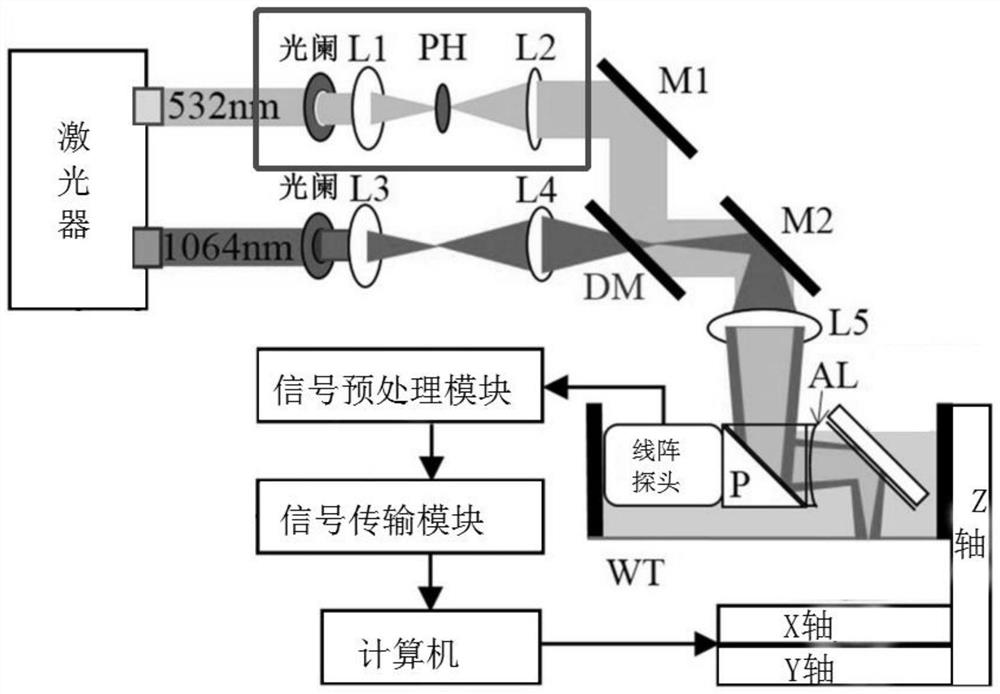
[0058] Such as Figure 4 As shown, the present invention also provides a photoacoustic imaging method for breast, comprising the following steps:

[0059] S1. Collect the photoacoustic signal generated after the laser irradiates the breast tissue;

[0060] S2. Preprocessing the photoacoustic signal into a digital signal;

[0061] S3. Denoising the digital signal obtained after the preprocessing, reconstructing the optical coefficient absorption distribution of the breast tissue, performing medical imaging on the breast tissue, and obtaining a photoacoustic image of the breast;

[0062] S4. Extracting the region of interest of the mammary photoacoustic image;

[0063] S5. Locate the mammary gland lesion point according to the region of interest in the mammary gland photoacoustic image.

[0064] In this embodiment, the digital signal obtained after preprocessing is denoised, and the optical coefficient absorption distribution of breast tissue is reconstructed by using a phase...


PUM


Abstract

The invention discloses a photoacoustic imaging method and system for a mammary gland. The method comprises the following steps of collecting a photoacoustic signal generated after a mammary gland tissue is irradiated by a laser; preprocessing the photoacoustic signal and converting the photoacoustic signal into a digital signal; denoising the digital signal obtained after preprocessing, reconstructing optical coefficient absorption distribution of the mammary gland tissue, conducting medical imaging on the mammary gland tissue, and obtaining a mammary gland photoacoustic image; extracting a region of interest of the mammary gland photoacoustic image; and according to the region of interest of the mammary gland photoacoustic image, positioning a mammary gland lesion point. According to the photoacoustic signal acquisition method and system, the optical coefficient absorption distribution of the mammary tissue is reconstructed by adopting a phase-controlled focusing algorithm, the mammary tissue is subjected to medical imaging, the photoacoustic image can be reconstructed by acquiring the photoacoustic signals in one direction through a detector, rotation around the biological tissue is not needed, the signal acquisition process is fast, the purpose of real-time imaging is achieved, and no vibration error is introduced.

Description

technical field [0001] The invention relates to the technical field of medical imaging, in particular to a photoacoustic imaging method and system for mammary glands. Background technique [0002] Medical imaging of the internal structure of biological tissue is an important means of research and analysis of biological tissue. Among them, photoacoustic imaging is a non-destructive medical imaging method developed in recent years. It has the advantages of both optical and ultrasonic imaging. By introducing pulsed laser light into biological tissue, the tissue will generate ultrasonic signals due to instantaneous thermal expansion, and the tissue can be obtained by detecting the ultrasonic signal. The light absorption information, and then reflect the biological tissue information. It has a wide range of applications. For example, breast imaging based on photoacoustic imaging can study the pathogenesis of breast cancer based on the imaging of breast tissue. [0003] In the p...

Claims


Application Information

Patent Timeline
IPC IPC(8): A61B5/00G06T7/00G06T7/11G06T7/12G06T7/33
CPCA61B5/0095A61B5/0091A61B5/7203A61B5/748A61B5/7225A61B5/725G06T7/0012G06T7/11G06T7/12G06T7/337G06T2207/10132G06T2207/30068G06T2207/30096
Inventor 杨帆郑传胜赵煌旋黄嘉范文亮
Owner XIEHE HOSPITAL ATTACHED TO TONGJI MEDICAL COLLEGE HUAZHONG SCI & TECH UNIV
Features
  • R&D
  • Intellectual Property
  • Life Sciences
  • Materials
  • Tech Scout
  • Unparalleled Data Quality
  • Higher Quality Content
  • 60% Fewer Hallucinations
Learn More