
EGFR gene mutation detection method and system based on chest CT image

A CT image and detection method technology, applied in the field of artificial intelligence and medical image analysis, can solve the problems of small application range and limited application, and achieve the effect of wide application range

Active Publication Date: 2021-06-25
CHINA JAPAN FRIENDSHIP HOSPITAL
View PDF7 Cites 4 Cited by
  • Summary
  • Abstract
  • Description
  • Claims
  • Application Information

AI Technical Summary

Problems solved by technology

At present, there are some methods for predicting the EGFR gene mutation status of lung cancer patients based on radiomics features on chest CT images, but the existing methods are only applicable to one type of CT images, so the scope of application is small and limited. clinical application

Method used


Image

  • EGFR gene mutation detection method and system based on chest CT image
  • EGFR gene mutation detection method and system based on chest CT image
  • EGFR gene mutation detection method and system based on chest CT image

Examples


experiment example 1

[0129] Experimental Example 1 Regression Model and Performance Evaluation of Different Target Image Metal Features Screening Method

[0130] As shown in Table 1, it is statistically statistical conditions in the inclusion in the present invention, as shown in Table 1, divided by the patient into a training group and a verification group. Training groups include 327 lung cancer patients. In the training group, each patient has done a CT image, including 167 people as a flat-sweep CT image (N-CT), 160 people correspond to enhance CT images (E-CT) . The verification group consists of 66 patients with lung cancer, and each patient per patient has done two CT images (N-CT & E-CT).

[0131] Table 1

[0132]

[0133] The EGFR gene mutation state of the patient in the training group and the verification group is shown in Table 1, and the mutant type indicates that the patient is a mutation of EGFR gene, and wild type indicates that the patient is EGFR gene mutation negative.

[0134]The...

experiment example 2

[0146] Experimental Example 2 The Construction of Normount and Its Performance Comparative Experiments from NECT-Model

[0147] Screening of clinical features and radiological characteristics for patients in the training group in Table 1, wherein the clinical features include age, gender, smoking history, pathological type and chronic obstructive pulmonary disease (Chronic obstructive pulmonarydisease, COPD), etc. Scholars include tumors, position, mass nedule and opacity, pulmonary metathetic change, bronchitis, bronchial expansion, emphysema, lymphadenopathy, pleural thickening and pleural effusion, tumor imaging characteristics Dibrillation, needle, cavitation and pleural contraction, interstitial pulmonary disease (ILD), etc.

[0148] All clinical features and radiological features are monitored to assess whether they can be used as a predictor of the EGFR gene mutation. Multi-factor analysis results in multi-factor analysis results as a target clinical characteristic and targ...


PUM


Abstract

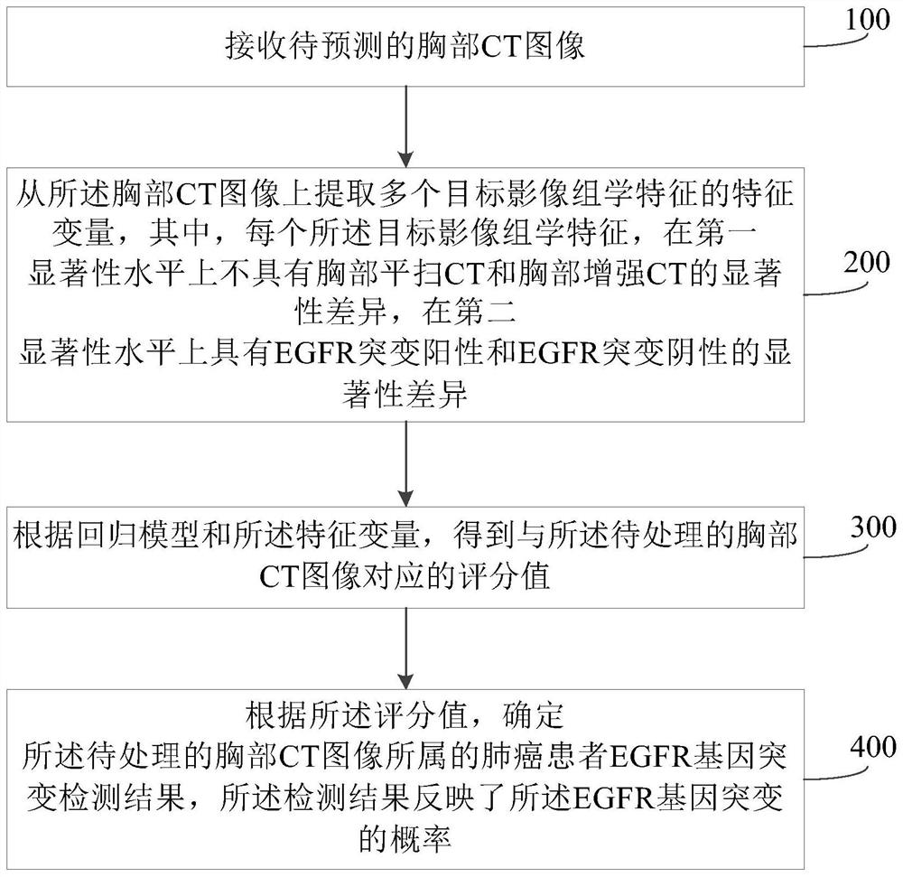
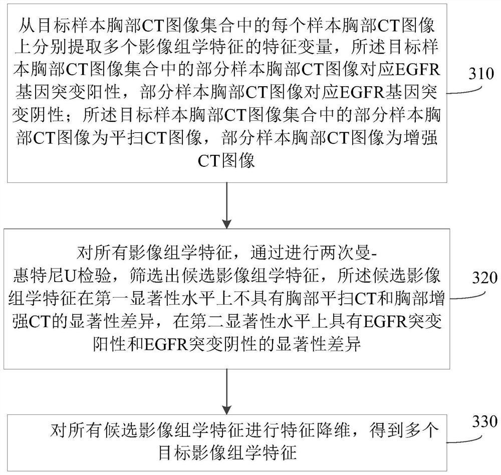
The invention discloses an EGFR gene mutation detection method and system based on a chest CT image and a computer readable storage medium. The method comprises the following steps: receiving a chest CT image to be processed; extracting feature variables of a plurality of target imageomics features from the chest CT image, wherein each target imageomics feature does not have a saliency difference between chest plain-scan CT and chest enhanced CT on a first saliency level and has a saliency difference between EGFR mutation positive and EGFR mutation negative on a second saliency level; according to a regression model and the feature variables, obtaining a score value corresponding to the chest CT image to be processed; according to the score value, determining an EGFR gene mutation detection result of a lung cancer patient to which the chest CT image to be processed belongs, wherein the detection result reflects the probability of EGFR gene mutation. According to the method, EGFR gene mutation can be detected based on a chest plain-scan CT image or an enhanced CT image, and the clinical application range is wide.

Description

Technical field [0001] The present invention relates to artificial intelligence and medical imaging analysis techniques, and more particularly to the EGFR gene mutation detection method, system, and computer readable storage medium based on a chest CT image. Background technique [0002] Lung Cancer (LC) is the most common malignant tumor in the lungs, and there are about 1.8 million people in the world. The incidence rate in the past 50 years is significantly increased. Lung cancer is clinically divided into two categories: Non-Small Celllung Cancer (NSCLC) in clinically, of which NSCLC accounts for 80%, and the most common NSCLC organizes subtype gland cancer and squamous cell carcinoma. Squamous Cell Carcinoma, SQCC). Epidermal Growth Factorreceptor, EGFR) tyrosine kinase inhibitor (TKIS) can increase the median survival of EGFR gene sensitive mutation lung cancer, and improve its living quality, and EGFR gene mutation is negative or Patients with non-sensitive mutations canno...

Claims


Application Information

Patent Timeline
IPC IPC(8): A61B6/03G06T7/00G06T7/11
CPCA61B6/032A61B6/52A61B6/5211G06T7/0012G06T7/11G06T2207/10081G06T2207/30061
Inventor 杨晓燕刘敏
Owner CHINA JAPAN FRIENDSHIP HOSPITAL
Learn More