
Foresight 3D endoscopic ultrasound system and imaging method

An ultrasound system and imaging method technology, applied in the field of clinical medicine, can solve the problems of high price, difficult to make small size, incapable of imaging and diagnosis and treatment, and achieve the effect of low cost and simple structure

Pending Publication Date: 2021-07-23
SUZHOU INST OF BIOMEDICAL ENG & TECH CHINESE ACADEMY OF SCI
View PDF0 Cites 0 Cited by
  • Summary
  • Abstract
  • Description
  • Claims
  • Application Information

AI Technical Summary

Problems solved by technology

[0005] 1) For components that require mechanical retraction, real-time 3D imaging cannot be performed, and only side-view images can be formed, but front-view images cannot be formed;
[0006] 2) For the electronic scanning method, it needs an area array probe, its size is difficult to make small, and the price is very high, while in vivo ultrasound imaging is limited by the volume of blood vessels, digestive tract, bronchi, etc., and most of them are disposable consumables , making it difficult to promote the app
[0007] However, the forward-looking 3D image has a very urgent clinical demand, such as chronic total occlusion (CTO) of coronary artery, or the presence of intraluminal thrombus, the imaging catheter cannot be passed through, and imaging, diagnosis and treatment cannot be performed.
At the same time, CT and other in vitro diagnostic methods cannot assess the specific severity because of the inability to angiography after obstruction

Method used


Image

  • Foresight 3D endoscopic ultrasound system and imaging method
  • Foresight 3D endoscopic ultrasound system and imaging method
  • Foresight 3D endoscopic ultrasound system and imaging method

Examples


Embodiment Construction

[0042] In order to make the above objects, features and advantages of the present invention more obvious and understandable, the present invention will be described in detail below in conjunction with the accompanying drawings and specific embodiments. In the following description, numerous specific details are set forth in order to provide a thorough understanding of the present invention. However, the present invention can be implemented in many other ways different from those described here, and those skilled in the art can make similar improvements without departing from the connotation of the present invention, so the present invention is not limited by the specific embodiments disclosed below.

[0043] In describing the present invention, it should be understood that the terms "center", "longitudinal", "transverse", "length", "width", "thickness", "upper", "lower", "front", " Back", "Left", "Right", "Vertical", "Horizontal", "Top", "Bottom", "Inner", "Outer", "Clockwise"...


PUM


Abstract

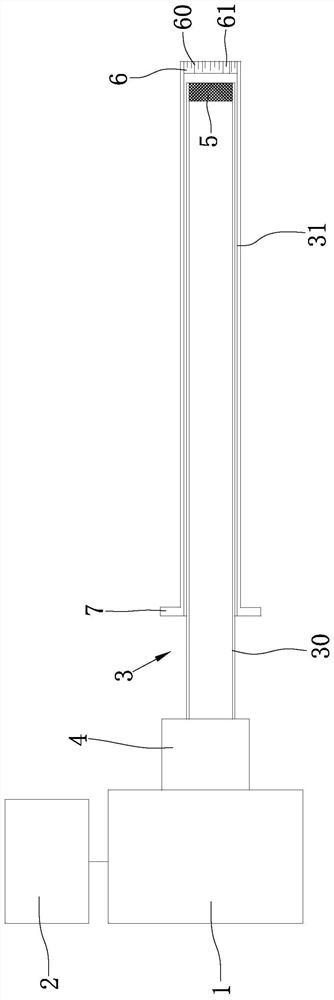
The invention relates to a foresight 3D endoscopic ultrasound system and an imaging method. The endoscopic ultrasound system comprises a host, a display, an endoscopic catheter and a communicating vessel; the endoscopic catheter comprises an inner catheter and an outer cannula which are arranged in a coaxial and relative rotation manner; the foresight 3D endoscopic ultrasonic system further comprises an ultrasonic transducer arranged at the front end of the inner catheter and an end plate arranged at the front end of the outer cannula; the working face of the ultrasonic transducer faces the end plate and is close to the inner wall of the end plate; stripes are formed on the end plate; and a transmission path of ultrasonic waves, emitted by the ultrasonic transducer, passing through a stripe area changes along with the change of the stripes, and phase delay is formed. According to the invention, through the arrangement of the internal catheter and the outer cannula which rotate relative to each other and the cooperation of the single ultrasonic transducer with the working face facing the stripes, data information of ultrasonic excitation and echo signal receiving is obtained under multiple relative rotation angles, so that a 3D image is accurately reconstructed; and the structure is simple, and the cost is low.

Description

technical field [0001] The invention belongs to the technical field of clinical medicine, and specifically relates to a forward-looking 3D endoscopic ultrasound system, and also relates to an imaging method of the forward-looking 3D endoscopic ultrasound system. Background technique [0002] As we all know, in vivo ultrasound imaging, which mainly includes intravascular ultrasound imaging, digestive endoscopic ultrasonography, and ultrasound bronchoscopy, has become an important tool for the diagnosis and auxiliary minimally invasive treatment of cardiovascular diseases, digestive and respiratory diseases. 3D imaging, on the other hand, can present the tissue structure in a more three-dimensional manner, and can provide more visual and intuitive information on vascular stenosis, plaque distribution, tumor lesion volume, and infiltration range, which has become a trend in the development of related products in recent years. [0003] However, at present, there are two main sch...

Claims


Application Information

Patent Timeline
Patent Type & Authority Applications(China)
IPC IPC(8): A61B8/12A61B8/08
CPCA61B8/12A61B8/483A61B8/4483A61B8/58A61B8/0891
Inventor 简小华韩志乐王宁浩
Owner SUZHOU INST OF BIOMEDICAL ENG & TECH CHINESE ACADEMY OF SCI
Learn More