
Cervical cell image screening method and system, computer equipment and storage medium

A technology of cervical cells and storage media, which is applied in computing, image enhancement, image analysis, etc., can solve problems such as cell division and deep learning model learning deviation, so as to improve accuracy, improve correct rate, and reduce labor intensity and workload Effect

Inactive Publication Date: 2021-08-06
SHENZHEN GRADUATE SCHOOL TSINGHUA UNIV
View PDF3 Cites 0 Cited by
  • Summary
  • Abstract
  • Description
  • Claims
  • Application Information

AI Technical Summary

Problems solved by technology

However, the sliding of fixed-size windows will inevitably lead to the following problems: 1. Too much invalid information is introduced, such as background, impurities, etc.; 2. Cells are split in different pictures; 3. The same picture may contain both negative and positive cells
These issues can bias the learning of deep learning models

Method used


Image

  • Cervical cell image screening method and system, computer equipment and storage medium
  • Cervical cell image screening method and system, computer equipment and storage medium
  • Cervical cell image screening method and system, computer equipment and storage medium

Examples

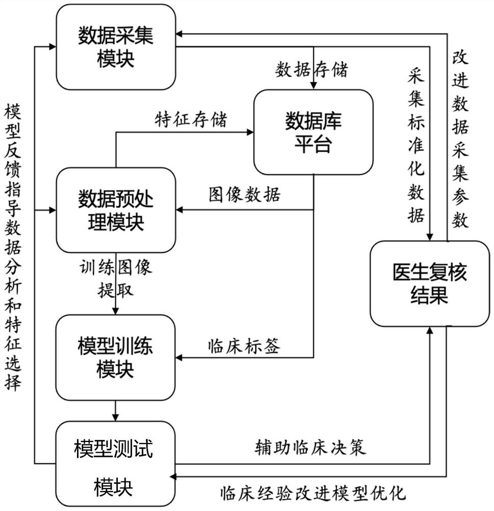

Embodiment Construction

[0025] The present invention will be described in further detail below in conjunction with specific embodiments and with reference to the accompanying drawings. It should be emphasized that the following descriptions are only exemplary and not intended to limit the scope of the present invention and its application.

[0026] In the previous cervical cell image screening process, the liquid-based cell slides were generally scanned by the instrument and then stored for the user. The user then went to manually read the slides or found an AI pathological diagnosis company for preliminary screening and auxiliary slide reading. However, in order to improve the accuracy of liquid-based cell screening and increase the speed of cell recognition, this embodiment directly develops a cervical cell image screening system on a liquid-based cell film production and scanning machine, and transmits the scanned original image to trigger an algorithm for assistance. Diagnosis, the user confirms t...


PUM


Abstract

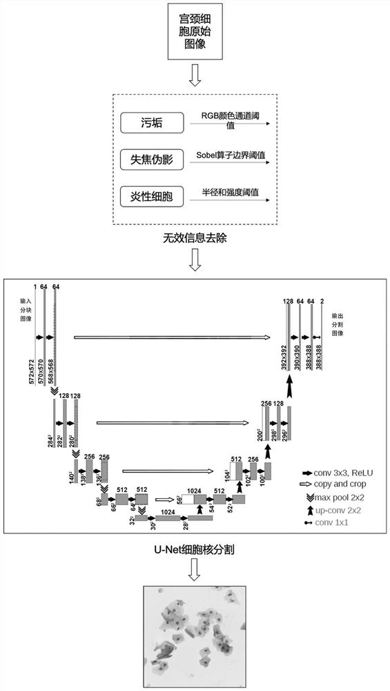
The invention provides a cervical cell image screening method and system, computer equipment and a storage medium. The method comprises the following steps: S1, preprocessing an original image containing cervical cells; S2, establishing a cervical cell image screening model and performing training; and S3, inputting the image preprocessed in the step S1 into the cervix uteri cell model established in the step S2 for screening the cervical cell image. Traditional manual radiograph reading is replaced, automatic and end-to-end cervical cell image screening is adopted, the diagnosis work efficiency can be effectively improved, the labor intensity and workload of pathologists are reduced, the influence of subjective factors in manual operation is avoided, and finally the accuracy is improved. In the step S1, invalid information removal is carried out on the original image, wherein the invalid information comprises dirt, out-of-focus artifacts and inflammatory cells; and then cell nucleus segmentation is carried out by using a U-Net structure automatic segmentation network, so that the accuracy of cell segmentation is improved.

Description

technical field [0001] The present invention relates to the technical field of artificial intelligence, in particular to a screening method, system, computer equipment and computer-readable storage medium for cervical cell images. Background technique [0002] According to statistics, 300,000 people die from cervical cancer every year in the world, and 450,000 are diagnosed and 450,000 with early symptoms. Although the age of diagnosed patients is generally over 35 years old, women with predisposing factors for this disease are often much younger than this age. Early cervical cancer is curable if diagnosed in time. However, because women in developing countries know less about this disease, coupled with medical and economic constraints, they often lose the opportunity of early diagnosis and treatment. Especially in the central and western rural areas of our country, due to the lack of funds and medical technology, cervical cancer is the enemy of women. Women in cities als...

Claims


Application Information

Patent Timeline
Patent Type & Authority Applications(China)
IPC IPC(8): G06T7/00
CPCG06T7/0012G06T2207/20081G06T2207/20084G06T2207/30096
Inventor 宋盟春胡梦颖黄强关添何永红
Owner SHENZHEN GRADUATE SCHOOL TSINGHUA UNIV
Learn More