Lung artery and vein segmentation method and device for CT image, medium and equipment

A CT image and image segmentation technology, applied in the field of medical image processing, can solve problems such as low segmentation accuracy, difficult segmentation of pulmonary artery and pulmonary vein, and difficulty in meeting clinical application requirements, achieving high accuracy and meeting clinical application requirements.

Pending Publication Date: 2021-09-17
NORTHEASTERN UNIV
View PDF10 Cites 2 Cited by
  • Summary
  • Abstract
  • Description
  • Claims
  • Application Information

AI Technical Summary

Problems solved by technology

The segmentation of pulmonary artery and pulmonary vein is of great significance to the detection of lung diseases. The segmentation of pulmonary artery and pulmonary vein in CT images is very difficult, because the dose of angiographic contrast agent and scanning time have a great influence on the results of angiography. Pulmonary vascular circulation is fast, and the angiographic results obtained by different doctors and equipment scans will have a relatively large difference in gray value. When the existing automatic segmentation method performs arteriovenous segmentation, it can only effectively segment on part of the data. , the segmentation accuracy on most of the data is low, and it is difficult to meet the needs of clinical applications

Method used


Image

  • Lung artery and vein segmentation method and device for CT image, medium and equipment
  • Lung artery and vein segmentation method and device for CT image, medium and equipment
  • Lung artery and vein segmentation method and device for CT image, medium and equipment

Examples


Embodiment 1

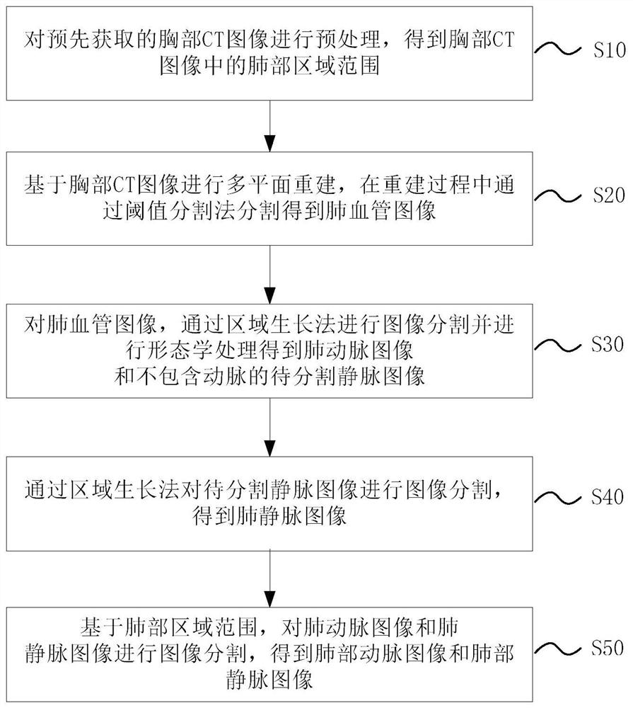
[0057] figure 1 It is a schematic flow chart of the pulmonary artery and vein segmentation method for CT images in one embodiment of the present application, as figure 1 As shown, the method includes:

[0058] S10. Preprocessing the pre-acquired chest CT image to obtain the range of the lung area in the chest CT image;

[0059] S20. Perform multi-planar reconstruction based on the chest CT image, and segment the pulmonary blood vessel image through the threshold segmentation method during the reconstruction process; wherein, the threshold used in the threshold segmentation method is the CT determined based on the two-dimensional segmentation result of the reconstructed lung region value range;

[0060] S30. For the pulmonary blood vessel image, select a pixel point from the right ventricle pulmonary artery area as the first seed point, select the first sub-interval from the CT value interval as the first threshold interval, perform image segmentation by region growing method...

Embodiment 2

[0115] Corresponding to the pulmonary artery and vein segmentation method of the above CT image, Figure 9 It is a schematic diagram of the structure of the pulmonary artery and vein segmentation device for CT images in another embodiment of the present application, refer to Figure 9 The apparatus 900 for pulmonary artery and vein segmentation of CT images includes: a preprocessing module 910 , a pulmonary vessel image segmentation module 920 , a pulmonary artery image segmentation module 930 , a pulmonary vein image segmentation module 940 and a pulmonary arteriovenous image segmentation module 950 .

[0116] A preprocessing module 910, configured to preprocess the pre-acquired chest CT image to obtain the lung area range in the chest CT image;

[0117] The pulmonary vessel image segmentation module 920 is configured to perform multi-planar reconstruction based on the chest CT image, and segment the pulmonary vessel image through threshold segmentation during the reconstruct...

Embodiment 3

[0124] The third aspect of the present application provides an electronic device through another embodiment, including: a memory, a processor, and a computer program stored on the memory and operable on the processor. When the computer program is executed by the processor, the above embodiments are realized. The steps of the pulmonary artery and vein segmentation method of the CT image described in any one.

[0125] Figure 10 It is a schematic structural diagram of an electronic device in another embodiment of the present application.

[0126] Figure 10 The illustrated electronic device may include: at least one processor 101 , at least one memory 102 , at least one network interface 104 and other user interfaces 103 . Various components in the electronic device are coupled together through the bus system 105 . It can be understood that the bus system 105 is used to realize connection and communication between these components. In addition to the data bus, the bus system...


PUM


Abstract

The embodiment of the invention relates to a lung artery and vein segmentation method and device for a CT image, a medium and electronic equipment, and the method comprises the steps: carrying out the preprocessing of a chest CT image obtained in advance, and obtaining a lung region range in the chest CT image; performing multi-plane reconstruction based on the chest CT image, and performing segmentation through a threshold segmentation method in the reconstruction process to obtain a pulmonary blood vessel image; carrying out image segmentation on the pulmonary blood vessel image through a region growing method to obtain a pulmonary artery image and a to-be-segmented vein image which does not contain arteries; performing image segmentation on the vein image to be segmented through a region growing method to obtain a pulmonary vein image; and based on the lung region range, carrying out image segmentation on the pulmonary artery image and the pulmonary vein image to obtain the pulmonary artery image and the pulmonary vein image. According to the lung artery and vein segmentation method, the segmentation result is high in accuracy, and the clinical application requirement can be met.

Description

technical field [0001] The present application belongs to the technical field of medical image processing, and in particular relates to a method, device, medium and equipment for pulmonary artery and vein segmentation of CT images. Background technique [0002] Pulmonary blood vessels are an independent blood circulation system, the main function is to transport blood to the lungs or take blood away from the lungs, so as to complete the exchange of blood in the lungs. Pulmonary blood vessel segmentation has been obtained in the field of biomedical image processing. Extensively studied, however, distinguishing between the pulmonary artery tree and the pulmonary vein tree remains a challenge. The segmentation of pulmonary artery and pulmonary vein is of great significance to the detection of lung diseases. The segmentation of pulmonary artery and pulmonary vein in CT images is very difficult, because the dose of angiographic contrast agent and scanning time have a great influe...

Claims


Application Information

Patent Timeline
Patent Type & Authority Applications(China)
IPC IPC(8): G06T7/11G06T7/136G06T7/155G06T7/187
CPCG06T7/11G06T7/136G06T7/155G06T7/187G06T2207/10081G06T2207/30061G06T2207/30101G06T2207/20036G06T2207/30204
Inventor 周庆华覃文军张子明李小硕杨金柱赵大哲
Owner NORTHEASTERN UNIV
Why Eureka
Try Eureka