Simple and effective method for induced amplification of iNKT cells and application

A NKT cell and cell technology, applied in animal cells, vertebrate cells, cell culture active agents, etc., can solve the problems of unsatisfactory anti-tumor effect, large demand for initial cells, and low remission rate.

Active Publication Date: 2021-10-01
XUZHOU MEDICAL UNIV
View PDF4 Cites 1 Cited by
  • Summary
  • Abstract
  • Description
  • Claims
  • Application Information

AI Technical Summary

Problems solved by technology

However, due to the low content of iNKT cells in human peripheral blood (accounting for about 0.01-1% of lymphocytes), the current expansion method has technical problems such as large d...

Method used


Image

  • Simple and effective method for induced amplification of iNKT cells and application
  • Simple and effective method for induced amplification of iNKT cells and application
  • Simple and effective method for induced amplification of iNKT cells and application

Examples


Embodiment 1

[0115] Example 1. Preparation of iNKT cells

[0116]1) Separation of PBMCs: collect peripheral blood from the donor, dilute the whole blood with an equal volume of normal saline, add the lymphocyte separation solution and the diluted blood to the centrifuge tube at a ratio of 1:2, centrifuge at 2000rpm / min for 20 minutes, and collect the buffy coat cells , washed twice with normal saline, and centrifuged at 1500 rpm / min for 8 minutes to obtain PBMCs from peripheral blood mononuclear cells.

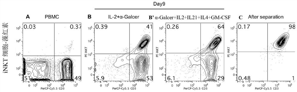
[0117] 2) Induce iNKT cells: resuspend PBMCs with lymphocyte medium, adjust the cell concentration to 2×10 6 / mL, add 100ng / mLα-Galcer, 50U / mL IL-2, 10ng / mL IL-21, 500U / mL IL-4 and 500U / mL GM-CSF, inoculate the cells in a 24-well plate and place at 37°C , 5% CO2 incubator. Observe the cell state every day, change the medium in half every other day, monitor the expansion effect by cell counting, and analyze the phenotype of iNKT cells by flow cytometry.

[0118] 3) Magnetic bead sortin...

Embodiment 2

[0126] Example 2. The activity of iNKT cells to kill tumor cells in vitro

[0127] Effector cells: iNKT cells obtained in Example 1

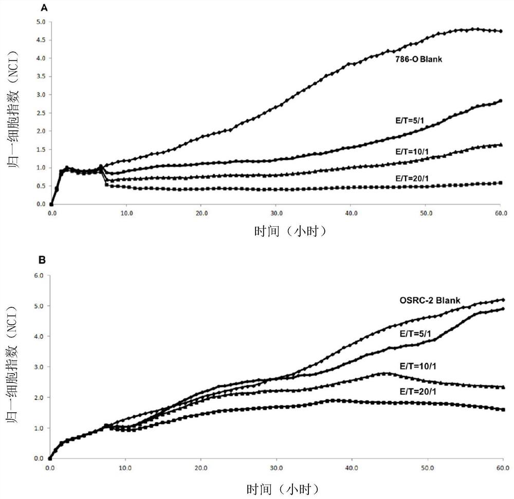
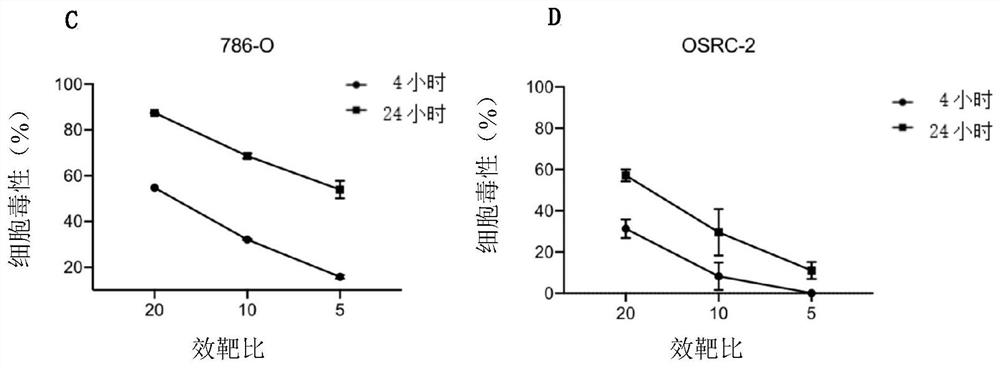
[0128] Target cells: 786-O cells and OSRC-2 cells (this example takes human renal cancer cells as an example)

[0129] First, add 50 μL of tumor cell complete medium to the E-Plate detection plate of the xCELLigence cell function analyzer, and measure the background impedance value; collect the target cells in the logarithmic phase, adjust the concentration of the cell suspension to 1×10⁵ / mL, and transfer to E -Add 100 μL of cell suspension to the plate, let it stand at room temperature for 30 minutes, and then place it on the detection platform; observe the real-time dynamic observation when the target cell proliferation is in the plateau stage, according to the effect-to-target ratio of 20:1, 10:1, 5:1 50 μL of effector cells were added, and only T cell culture medium was added to the control group; continue to observe the cell killing effe...

Embodiment 3

[0134] Example 3. Ability of iNKT cells to secrete cytokines

[0135] Digest tumor cells, wash and count to adjust the density to 1×10 5 / mL, pave a 24-well plate, and adhere to the wall overnight at 2 mL per well; collect the expanded iNKT cells, add them to the tumor cells at a ratio of 1:1 for co-cultivation, collect the supernatant after 24 hours, and detect IFN-γ and IL- 2. TNF-α.

[0136] image 3 The results showed that co-incubation of iNKT cells and renal cancer cells could secrete cytokines A.IFN-γ, B.IL-2 and C.TNF-α.

[0137] The above results prove that the iNKT cells obtained in Example 1 have the ability to secrete cytokines.


PUM


Abstract

The invention provides a method for induced amplification of iNKT cells. According to the method, the initial demand quantity is small (-10<6> orders of magnitude), the proportion of the obtained iNKT cells is high, the proportion of the iNKT cells is increased to 20-80% from 0.01-1% after 7-9 days, the efficiency can be remarkably improved, and the final amplification of the iNKT cells exceeds 2000 times; and the prepared cells can secrete Th1 type cytokines, highly express CD62L, have long in-vivo survival time and can meet the requirements of clinical treatment.

Description

technical field [0001] The invention belongs to the field of cellular immunotherapy and relates to a simple and effective method for inducing and expanding iNKT cells and its application. Background technique [0002] iNKT cells are named for their unique expression of invariant TCR, which mainly recognize glycolipid antigens presented by CD1d molecules, secrete a large number of cytokines, and mediate innate immunity and adaptive immunity at the same time. At present, iNKT cells are considered to be a group of rare T cells with powerful immune regulation and effector functions, and play an important role in anti-tumor immunity. [0003] At present, many studies have shown that there are low iNKT cell content or impaired function (reduced ability to secrete IFN-γ) in tumor patients. The number of infiltrating iNKT cells in the tumor is closely related to the clinical prognosis of neuroblastoma and colon cancer; a study with a median follow-up time of 8.7 years reported that...

Claims


Application Information

Patent Timeline
IPC IPC(8): C12N5/0783A61K35/17A61P35/00
CPCC12N5/0646A61K35/17A61P35/00C12N2501/2307C12N2501/2315C12N2501/999C12N2501/2302C12N2501/2321C12N2501/2304C12N2501/22C12N2501/515C12N2501/51
Inventor 李慧忠郑骏年王刚刘宜林张连军
Owner XUZHOU MEDICAL UNIV
Why Eureka
Try Eureka