
3D gradient spin echo diffusion imaging method for inversion recovery preparation, medium and equipment
A technology of inversion recovery and spin echo used in 3D modeling, medical imaging, image data processing, etc.
Patent Information
- Authority / Receiving Office
- CN · China
- Current Assignee / Owner
- Publication Date
- 2021-10-08
Smart Images
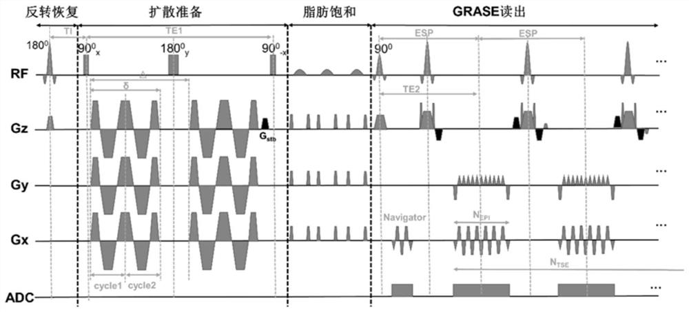
Figure 1 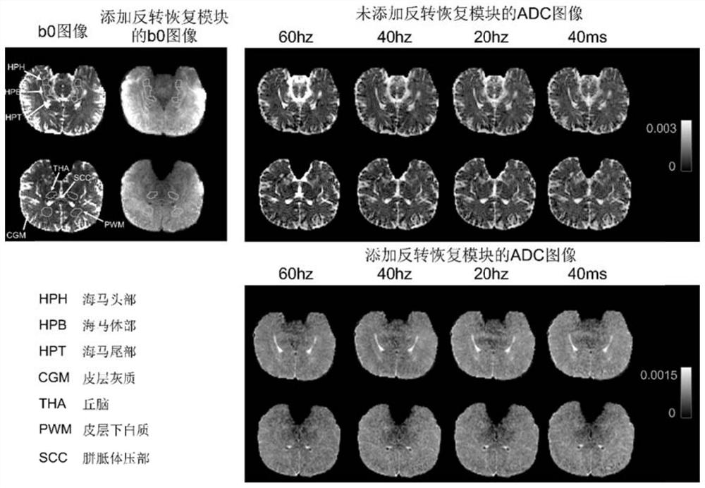
Figure 2 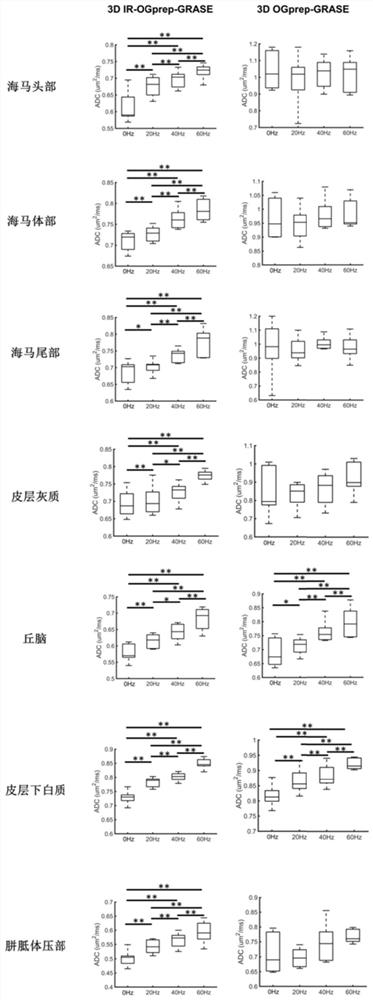
Figure 3
Abstract
Description
technical field
[0001] The invention relates to the field of magnetic resonance technology, in particular to the field of diffusion magnetic resonance imaging. Background technique
[0002] Diffusion magnetic resonance (dMRI) reveals various details of tissue microstructure by detecting the diffusion of water confined by the microstructural environment. Therefore, the apparent diffusion coefficient (ADC) measured by dMRI usually depends on the diffusion time of water molecules in the surrounding environment (t d ). A series of t d Diffusion of water molecules in the range (milliseconds to tens of milliseconds), and infer tissue microstructure, has been widely used in t d Dependent dMRI studies.
[0003] However, due to the low gradient strength of clinical MRI systems, longer echo times and repetition times are required to achieve reasonable oscillation frequencies and b-values. This leads to issues such as limited ability to access high-frequency oscillatory gradients,...
Examples
Embodiment
[0042] The above-mentioned 3D gradient spin echo diffusion imaging method prepared by inversion recovery, that is, the corresponding 3DIR-OGprep-GRASE sequence, and the 3D gradient spin echo diffusion imaging method without an inversion recovery module, that is, the corresponding 3D OGprep-GRASE sequence in 6 The tests were carried out in three healthy young male volunteers, and the specific parameters here are described below: The magnetic resonance scans used a Siemens Prisma 3T scanner (maximum gradient 80mT / m, maximum switching rate 200mT / m), and all scans were performed using 64 channel head coil.
[0043] Experiment: To compare 3D OGprep-GRASE and 3D IR-OGprep-GRASE sequences on diffusion information in the vicinity of CSF d Accuracy of dependent measurements, scans performed using pulsed diffusion gradient (0 Hz) and oscillating diffusion ladders at 20 Hz, 40 Hz, 60 Hz, other imaging parameters identical: Diffusion weighting = 420 s / mm 2 , 6 directions, 2 repetitions, ...