
Multiplane Microscopic Imaging System Based on Gradient Variable Index Lens

A gradient variable refractive index and microscopic imaging technology, which is applied in microscopes, instruments, optics, etc., can solve the problems of inability to obtain information on the fine structure of brain tissue, inability to image, and limited imaging speed, etc., to achieve rapid multi-plane imaging at any depth The effect of microscopic imaging

Active Publication Date: 2022-01-18
TSINGHUA UNIV
View PDF6 Cites 0 Cited by
  • Summary
  • Abstract
  • Description
  • Claims
  • Application Information

AI Technical Summary

Problems solved by technology

However, the traditional optical microscopy technology can only clearly image the tissue structure at the focal plane in a single shot, and cannot image objects outside the focal plane.
In addition, traditional optical microscopy techniques are affected by brain tissue scattering and cannot image tissues below 300 μm
The above two shortcomings greatly limit the application of optical microscopy in brain imaging
[0003] In related technologies, in order to realize the observation of multi-plane samples, the following three schemes are usually adopted: the first one uses a mechanical device to move the objective lens to achieve different depth positions, but the mechanical inertia limits the imaging speed; the second one uses an electrically adjustable zoom lens Changing the focal length of the objective lens, this method improves the imaging speed compared with the first method, but is limited by the principle of the electrically adjustable lens, and the field of view is limited; the third method is the light field microscopic imaging method, by sacrificing the lateral resolution of the imaging system To realize the acquisition and reconstruction of the three-dimensional light field, not only the information of the fine structure of the brain tissue cannot be obtained, but also a large amount of data algorithms and time are needed to realize the three-dimensional reconstruction, and it is impossible to observe the brain tissue in real time
In addition, in order to achieve deep imaging, confocal microscopy and multiphoton scanning microscopy are the most commonly used methods at present, but the acquisition of images by scanning greatly reduces the time resolution of the system, and this problem is more serious in large field of view imaging

Method used


Image

  • Multiplane Microscopic Imaging System Based on Gradient Variable Index Lens
  • Multiplane Microscopic Imaging System Based on Gradient Variable Index Lens
  • Multiplane Microscopic Imaging System Based on Gradient Variable Index Lens

Examples


specific Embodiment 1

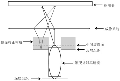
[0060] figure 2 According to an embodiment of the present invention, it is a principle diagram of an arbitrary-depth multi-plane wide-field microscopic imaging light based on a gradient variable refractive index lens, which is suitable for realizing multi-plane imaging for simultaneous observation of the hippocampus and cortical surface, and the gradient variable refraction The scene where the length of the power lens is greater than the depth of the tissue in the observation area.

[0061] Specifically, such as figure 2 As shown, the deep hippocampus tissue is mapped to the virtual image plane through the gradient variable refractive index lens, and the shallow tissue is also mapped to the same virtual image plane through the BK7 glass column. Such as image 3 As shown, the observation depth from the superficial layer of the brain tissue to the deep hippocampus is 1.5 mm. The gradient index lens has a diameter of 0.5mm and a length of 2mm. The cover glass has a diameter...

specific Embodiment 2

[0063] Figure 4 It is a principle diagram of another kind of arbitrary depth multi-plane wide-field microscopic imaging light based on gradient variable refractive index lens provided according to an embodiment of the present invention, which is suitable for realizing multi-plane imaging of simultaneous observation of hippocampus and cortical surface, and the gradient changes Scenarios in which the length of the refractive index lens is less than the depth of the tissue in the observation area.

[0064] Specifically, such as Figure 4 As shown, the deep hippocampus tissue is mapped to the virtual image plane 1 through the gradient variable refractive index lens and then mapped to the virtual image plane 2 through the BK7 glass column. At this time, the position of the virtual image plane 2 is consistent with that of the cortex. Such as Figure 5 As shown, the observation depth of the brain tissue from the superficial layer to the deep hippocampus was 1.5 mm. The gradient ...


PUM


Abstract

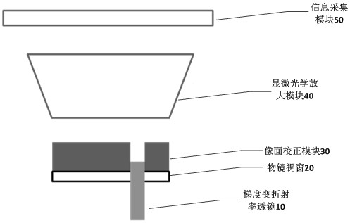
The invention discloses an arbitrary-depth multi-plane microscopic imaging system and method based on a gradient variable refractive index lens. The system includes: a gradient variable refractive index lens for generating an observation area of ​​the gradient variable refractive index lens on the focal plane of the microscope The virtual image of the sample to be imaged within the image plane correction module, which is used to generate the virtual image of the sample to be imaged outside the observation area of ​​the gradient variable refractive index lens, and adjust the position of the image plane outside the observation area of ​​the gradient variable refractive index lens; The micro-optical amplification module is used to collect the optical signal from the focal plane of the microscope and perform optical amplification; the information acquisition module is used to receive the optical signal output by the micro-optical amplification module and perform imaging. This system enables the traditional optical microscope to realize the simultaneous observation of the surface information of the observation sample, any deep layer or section information, improves the imaging speed, and expands the imageable depth area.

Description

technical field [0001] The invention relates to the field of optical microscopic imaging, in particular to an arbitrary-depth multi-plane microscopic imaging system and method based on a gradient variable refractive index lens. Background technique [0002] Optical microscopy imaging technology is a technology to obtain high-resolution images of surrounding objects through optical methods. It is currently widely used in structural imaging and functional signal detection of cells, bacteria, viruses and other microorganisms, and has become a common method in biological research. Among them, in the research of brain science, combined with chemical indicators, optical microscopy can detect changes in neuron activity by using changes in fluorescence intensity caused by changes in calcium concentration, chemical transmitter concentrations, and voltage changes. It is understandable that the brain has a layered three-dimensional structure, different layers have different structures ...

Claims


Application Information

Patent Timeline
Patent Type & Authority Patents(China)
IPC IPC(8): G02B21/00
CPCG02B21/00
Inventor 谢浩庄超玮贺敬戴琼海
Owner TSINGHUA UNIV
Learn More