Automatic ventricular septum dithering detection system based on ultrasonic image

An automatic detection and ultrasonic image technology, applied in the field of computer vision, can solve problems such as poor repeatability, investment in learning time cost, and failure to meet clinical needs well, and achieve the effect of simple method, fast calculation speed, and avoiding difference in results

Pending Publication Date: 2021-10-29
NORTHEASTERN UNIV +1
View PDF0 Cites 0 Cited by
  • Summary
  • Abstract
  • Description
  • Claims
  • Application Information

AI Technical Summary

Problems solved by technology

This method requires doctors to combine ultrasound images with LVGLS and other parameters, and the evaluation results depend on the doctor's clinical experience and the accuracy of LVGLS, and because there is no uniform standard for LVGLS, the repeatability of the experiment is poor
The parameters required for the experiment need to invest in learning, labor and time costs, which cannot well meet the clinical needs

Method used


Image

  • Automatic ventricular septum dithering detection system based on ultrasonic image
  • Automatic ventricular septum dithering detection system based on ultrasonic image
  • Automatic ventricular septum dithering detection system based on ultrasonic image

Examples


Embodiment Construction

[0024] The specific implementation manners of the present invention will be further described in detail below in conjunction with the accompanying drawings and embodiments. The following examples are used to illustrate the present invention, but are not intended to limit the scope of the present invention.

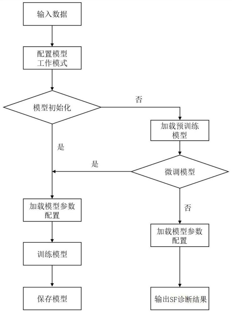
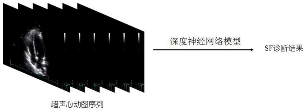
[0025] In this embodiment, a system for automatic detection of interventricular septum vibration based on ultrasound images, such as figure 1 , 2 As shown, the automatic detection of interventricular septal vibration is realized through the following steps:

[0026] Step 1: Acquire multiple echocardiograms as a sample dataset; based on the echocardiogram Figure four Cardiac view, to obtain echocardiogram files in dcm format following the Digital Imaging and Communications in Medicine (DICOM), or single-frame images (JPG, PNG, JPEG) after parsing echocardiogram files in dcm format format) and its corresponding diagnostic result label, that is, whether it is accompanied ...


PUM


Abstract

The invention provides an automatic ventricular septum jitter detection system based on an ultrasonic image, and relates to the technical field of computer vision. The system firstly obtains a plurality of echocardiograms with SF labels as a sample data set; then initializing a deep neural network model for ventricular septum jitter detection, and pre-training the deep neural network model by using the sample data set to obtain a pre-trained deep neural network model, wherein the deep neural network model used for ventricular septum jitter detection comprises a left ventricular segmentation network U-Net and a codec-based SF diagnosis network; and finally, loading model parameters and a configuration file of the pre-trained deep neural network model, segmenting the left ventricle in the echocardiogram to be evaluated, and outputting a ventricular septum jitter judgment result. According to the detection system, a deep learning method is used for automatically segmenting the left ventricle, automatic diagnosis of the SF is achieved according to the segmentation result, the clinical diagnosis time is shortened, and result differences caused by subjective experience of doctors are avoided.

Description

technical field [0001] The invention relates to the technical field of computer vision, in particular to an automatic detection system for interventricular septum shaking based on ultrasonic images. Background technique [0002] In patients with complete left bundle branch block (cLBBB) during early systole, the ventricular septum moves to the left followed by a reverse motion known as septal fluttering (SF). Patients with cLBBB who also have SF phenomena usually have poorer outcomes than those without SF phenomena. Usually, clinically, it is usually necessary to analyze and evaluate the phenomenon of ventricular septal vibration by combining the Left Ventricular Global Longitudinal Strain (LVGLS) and the original echocardiographic image. However, this method needs to trace the endocardium of the left ventricle, and the evaluation accuracy is highly dependent on the image quality and the doctor's experience level. Doctors with different experience levels often give differen...

Claims


Application Information

Patent Timeline
Patent Type & Authority Applications(China)
IPC IPC(8): G06T7/00G06T7/10G06K9/62G06N3/04
CPCG06T7/0012G06T7/10G06T2207/10132G06T2207/20081G06N3/045G06F18/214
Inventor 杨金柱马春燕瞿明军李洪赫王永槐曹鹏冯朝路覃文军栗伟
Owner NORTHEASTERN UNIV
Why Eureka
Try Eureka