Multi-knee-bending-angle knee joint finite element model and preparation method thereof

A technique of knee joint and multi-bend knee, which is applied in the finite element model of knee joint with multi-bend knee angle and its preparation field, achieves the effect of strong popularization, wide application range, and not cumbersome operation

Inactive Publication Date: 2021-12-31
FIRST AFFILIATED HOSPITAL OF KUNMING MEDICAL UNIV
View PDF0 Cites 0 Cited by
  • Summary
  • Abstract
  • Description
  • Claims
  • Application Information

AI Technical Summary

Problems solved by technology

At present, there is no finite element model of the knee joint with multiple knee flexion angles, whi...

Method used


Image

  • Multi-knee-bending-angle knee joint finite element model and preparation method thereof
  • Multi-knee-bending-angle knee joint finite element model and preparation method thereof
  • Multi-knee-bending-angle knee joint finite element model and preparation method thereof

Examples


Embodiment 1

[0034] A finite element model of a knee joint with multiple knee flexion angles and a preparation method thereof, comprising the following steps:

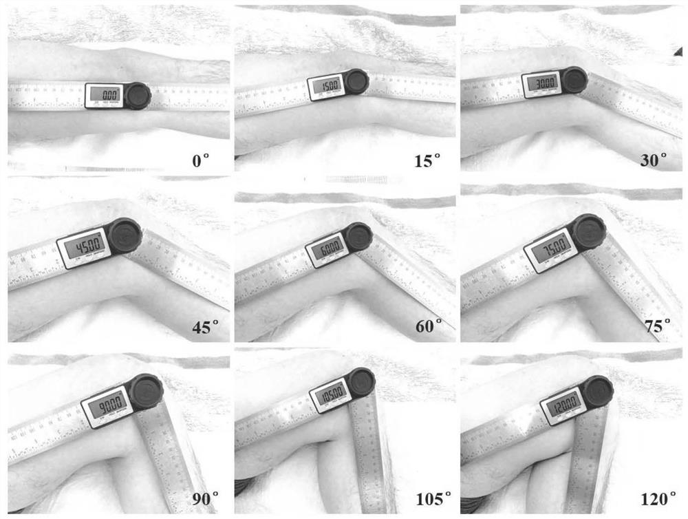
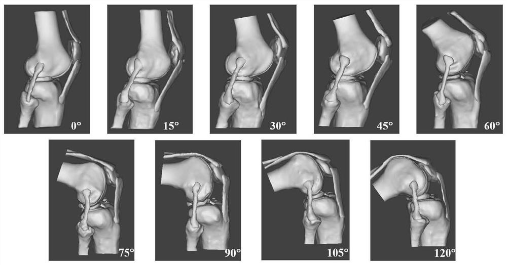
[0035] S1: MRI scans of healthy adult knee joints at multiple knee flexion angles: 0°, 15°, 30°, 45°, 60°, 75°, 90°, 105° and 120°. Use a two-in-one digital display angle ruler to adjust and fix the corresponding angles respectively, place it on the lower limb of the scanned side, and adjust the angle of the lower limb so that the long axis of the femur coincides with one end of the ruler, and the long axis of the tibia coincides with the other end of the ruler. Import the MRI data of multiple knee flexion angles into Mimics Research 19.0 software, and use "3D LiveWire" to reconstruct the femur, tibia, fibula, patella, femoral cartilage, medial tibial plateau cartilage, lateral tibial plateau cartilage, patellar cartilage, medial Meniscus, lateral meniscus, anterior cruciate ligament, posterior cruciate ligament, medial collateral ...

Embodiment 2

[0061] 1 Obtain the two-dimensional data of the knee joint

[0062] Multi-angle MRI scans of both knee joints were performed on healthy adults before operation ( figure 1 ), store the image in DICOM format on the workstation and burn it to CD-ROM, MRI parameters: 1.5-T superconducting nuclear magnetic resonance scanner (SIEMENS, Germany), using 3D-FSE (three-dimensional - fast spin echo) Sequence, specific scanning parameters: TR (repetition time): 1000ms, TE (echo time): 30ms, Voxel size (voxel): 0.6×0.6×0.9mm, FoV read (field of view): 160×160mm, FoV phase ( Rectangular field of view): 100%, Slice thickness (layer thickness): 0.9mm, Base resolution (matrix): 256×256, scanning time: 6min.

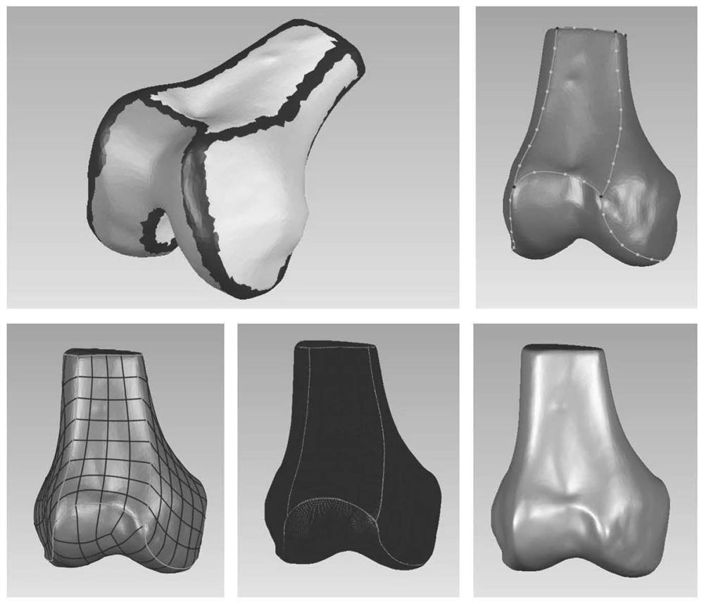
[0063] 2 Reconstruction of 3D digital model of human knee joint

[0064] On the computer workstation, the knee joint scanning image (DICOM) was imported into the interactive medical image control system Mimics Research 19.0 (Materialise, Belgium), and the femur, tibia, fibula, patella, a...


PUM


Abstract

The invention relates to a multi-knee-bending-angle knee joint finite element model and a preparation method thereof, and belongs to the field of joint anatomy. The model reversely reconstructs the main structure of the knee joint according to MRI data of healthy adults. The model collects and reconstructs a healthy knee joint three-dimensional model when actively bending the knee at 0 degree, 15 degrees, 30 degrees, 45 degrees, 60 degrees, 75 degrees, 90 degrees, 105 degrees and 120 degrees. The model comprises sixteen parts including a femur, a tibia, a fibula, a patella, a femur cartilage, a tibial plateau medial cartilage, a tibial plateau lateral cartilage, a patella cartilage, a medial meniscus, a lateral meniscus, an anterior cruciate ligament, a posterior cruciate ligament, a medial collateral ligament, a lateral collateral ligament, a quadriceps femoris tendon and a patellar tendon. The multi-knee-bending-angle knee joint finite element model is based on a real human knee joint, is complete in structure, is suitable for anatomical learning of the knee joint, and provides a unwonted real knee joint bending and stretching prediction model for simulating the actual condition of the knee joint, researching an injury mechanism, formulating a preoperative plan, clinical teaching and the like.

Description

technical field [0001] The invention belongs to the field of joint anatomy, and in particular relates to a finite element model of a knee joint with multiple knee flexion angles and a preparation method thereof. Background technique [0002] The knee joint is the largest and most complex weight-bearing joint in the human body. In recent years, the number of people participating in sports has continued to expand, and the incidence of knee joint sports injuries has gradually increased. With the introduction of precision medicine, more accurate biomechanical models are needed to study and analyze different structural injuries of the knee joint. At present, finite element analysis (FEA) is the most common research on knee biomechanics in clinical practice. Finite Element Analysis (FEA) uses mathematical approximation to simulate real geometric objects. By analyzing simple and interacting small units, namely A finite number of unknowns can be used to simulate the real situation...

Claims


Application Information

Patent Timeline
IPC IPC(8): G06F30/23G16H30/20G16H50/50
CPCG06F30/23G16H30/20G16H50/50
Inventor 李彦林刘德建杨贤光贾笛
Owner FIRST AFFILIATED HOSPITAL OF KUNMING MEDICAL UNIV
Why Eureka
Try Eureka