Multi-nuclide magnetic resonance multi-scale image registration method
A magnetic resonance image and image registration technology, which is applied in the field of medical image processing, achieves the effect of simple calculation method and reduced error
- Summary
- Abstract
- Description
- Claims
- Application Information
AI Technical Summary
Problems solved by technology
Method used
Image
Examples
Embodiment 1
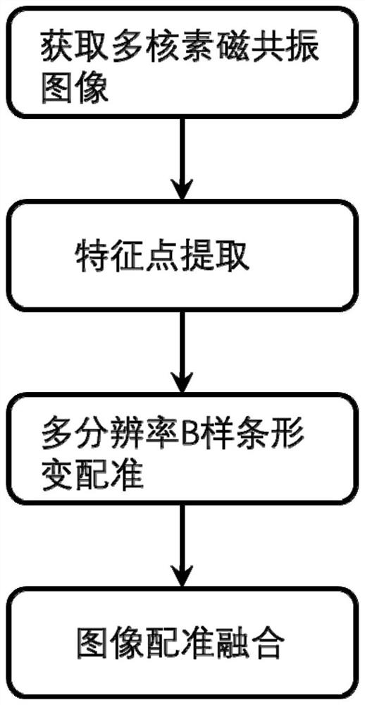
[0022] like figure 1 As shown, a multi-parentin magnetic resonance multi-scale image registration method, including the following steps:
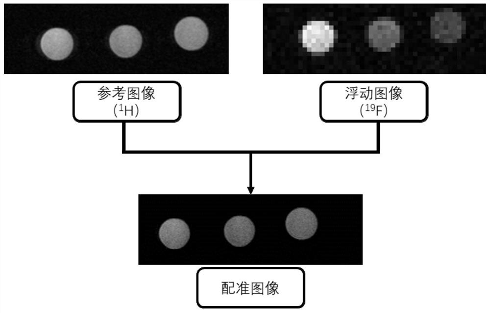
[0023] Step 1: Using a multinuclein synchronously integrated imaging magnetic resonance system, including including 1 H, 19 F, 23 NA, 31 Pyrovination of magnetic resonance images for collecting; the multiklane synchronous integrated imaging magnetic resonance system using patented CN107329100B;
[0024] Step 2: Select 1 H Magnetic resonance image as a reference image, 19 F magnetic resonance image as a floating image;
[0025] Step 3: Extract the reference image and the floating image, the feature point extraction includes the following steps: 1 H, 19 The F magnetic resonance image performs pyramid segmentation, using adjacent scale Gaussian filter smooth images, and further derives the processed image, reduce the image to the previous 1 / 4; repeat the above process, obtain a series of divisions Net structure;
[0026] Step 4: The reference imag...
Embodiment 2
[0028] A multi-parentin magnetic resonance multi-scale image registration method, including the following steps:
[0029] Step 1: Using a multinuclein synchronously integrated imaging magnetic resonance system, including 1 H, 19 F, 23 NA, 31 P and other magnetic resonance images are collected;
[0030] Step 2: Select 1 H Magnetic resonance image as a reference image, 23 Na magnetic resonance image as a floating image;
[0031] Step 3: Extract the reference image and the floating image, the feature point extraction includes the following steps: 1 H, 23 NA magnetic resonance image performs pyramid segmentation, using adjacent scale Gaussian filter smooth images, and further depends on the processed image, minimizing the image is previously 1 / 4, and repeats the above process, obtain a series of sections After the mesh structure;
[0032] Step 4: The reference image and the floating image are image registration by using the B-spline deformation method. The specific steps are as follow...
Embodiment 3
[0034] A multi-parentin magnetic resonance multi-scale image registration method, including the following steps:
[0035] Step 1: Using a multinuclein synchronously integrated imaging magnetic resonance system, including 1 H, 19 F, 23 NA, 31 P and other magnetic resonance images are collected;
[0036] Step 2: Select 1 H Magnetic resonance image as a reference image, 31 P magnetic resonance image as a floating image;
[0037] Step 3: Extract the reference image and the floating image, the feature point extraction includes the following steps: 1 H, 31 The P magnetic resonance image performs pyramid segmentation, using adjacent scale Gaussian filter smooth images, and further derives the processed image, minimize the image to 1 / 4, and repeat the above process multiple times, obtain a series of sections After the mesh structure;
[0038] Step 4: The reference image and the floating image are image registration by using the B-spline deformation method. The specific steps are as follow...
PUM
 Abstract
Description
Claims
Application Information
 - R&D
- Intellectual Property
- Life Sciences
- Materials
- Tech Scout
- Unparalleled Data Quality
- Higher Quality Content
- 60% Fewer Hallucinations
Browse by: Latest US Patents, China's latest patents, Technical Efficacy Thesaurus, Application Domain, Technology Topic, Popular Technical Reports.
© 2025 PatSnap. All rights reserved.Legal|Privacy policy|Modern Slavery Act Transparency Statement|Sitemap|About US| Contact US: help@patsnap.com


