Device for long-time living body imaging of waking small animals

A technology for live imaging and small animals, which is applied in the field of live imaging devices and devices for long-term live imaging of awake small animals. It can solve the problems that small animals cannot move freely, the imaging focal plane is unstable, and the bonding part is loose.

Pending Publication Date: 2022-03-22
ZHEJIANG UNIV
View PDF12 Cites 1 Cited by
  • Summary
  • Abstract
  • Description
  • Claims
  • Application Information

AI Technical Summary

Problems solved by technology

However, in fact, the real physiological state and changes of small animals can be observed and recorded more accurately only in the state of wakefulness and free movement. Due to the limitations of existing research devices, the observation of small animals in the state of wakefulness and free movement is two-photon A big problem in microscopy imaging
Common animal fixation devices are mostly used for imaging of small animals under anesthesia. Because of the existing fixation devices, small animals are generally glued to the imaging platform by dental cement, and small animals cannot move freely, and their limbs often only rest on the bottom plate. When the small animal is struggling and moving, it will have a reaction force on the bonding part, which will easily cause the bonding part to loosen and cause a large vibration, resulting in instability of the imaging focal plane. Therefore, it is usually necessary to adjust the Anesthetize the animal and keep it still
Existing methods are difficult to achieve long-term dynamic observation of small animals in an awake and free-moving state

Method used


Image

  • Device for long-time living body imaging of waking small animals
  • Device for long-time living body imaging of waking small animals
  • Device for long-time living body imaging of waking small animals

Examples


Embodiment 1

[0046] Example 1: Imaging of mouse brain in awake state

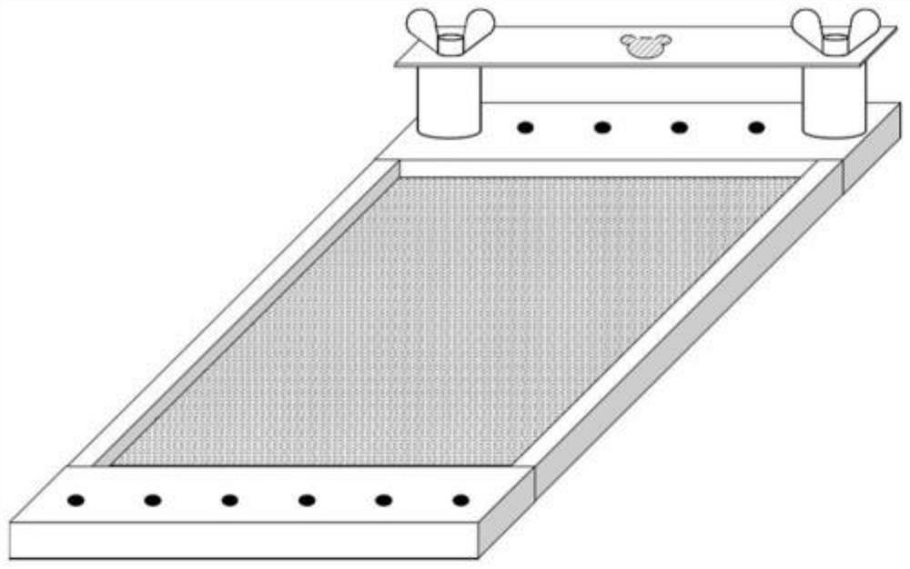
[0047] 1) Cut the scalp of the transgenic fluorescent mouse with ophthalmic scissors to expose the skull in the imaging area and stimulation administration area, treat the skull surface with hydrogen peroxide or ferric chloride solution, and remove the fascia; use dental cement (glass ionomer cement) The skull is fixed on the imaging fixed platform 2, and the imaging area and the stimulation and signal recording area are aligned with the imaging opening 4-1 and the stimulation and signal recording hole 4-2 of the platform.

[0048] 2) After the dental cement is completely solidified, according to the height of the head of the mouse, select a pillar 5 with a height of 2 cm and fix it in the screw hole on the bottom plate 6, and fix the imaging fixing platform 2 and the mouse together with the butterfly screw 1 on pillar 5.

[0049] 3) Use a bone drill to make holes in the imaging area and the stimulation and signal reco...

Embodiment 2

[0055] combine Figure 1-6 As shown, the difference from Example 1 is that the bottom plate 6 is a rectangular aluminum material, and M6 screw holes with a spacing of 25mm are distributed on the front and rear sides. It is used to fix the bottom plate on the microscope object platform or the microscope anti-vibration table, and can also be used to fix the pillar 5 of the imaging platform; as a further optimization, the pillar 5 can be provided with three pairs, which are 1cm, 2cm, and 3cm respectively. With cooperation, different height combinations from 1cm to 6cm, or at intervals of 1cm can be realized, so that it can be applied to small animals of different sizes;

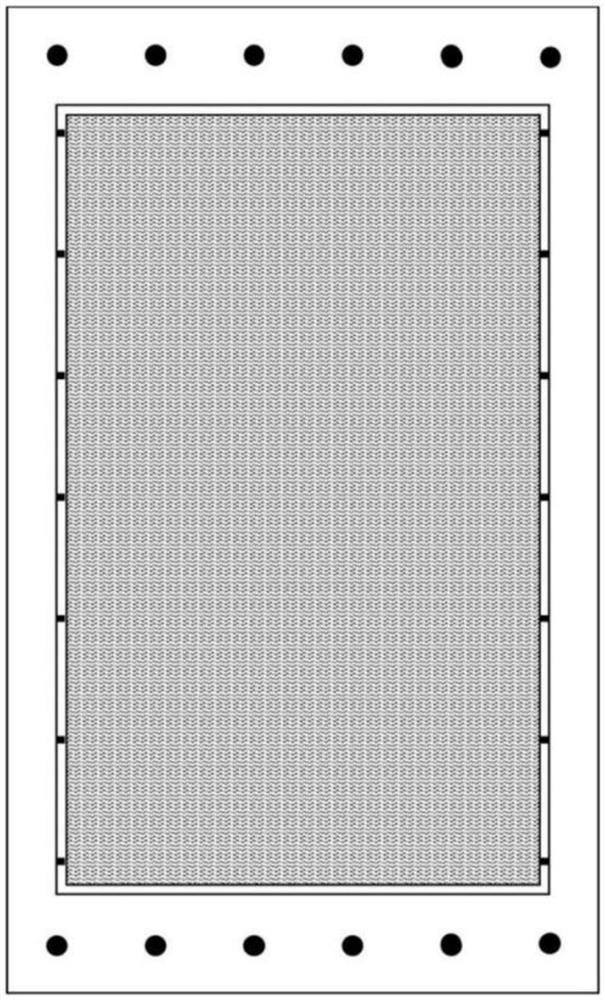
[0056] Both sides of described bottom plate 6 are all provided with fixing hole 15 and arc hole 9, and the center of circle of virtual circle where described arc hole 9 is located coincides with the center of circle of fixing hole 15, and described fixing hole 8 and arc hole 9 all use The movable crawler belt 7...


PUM


Abstract

The invention discloses a device and method for long-time living body imaging of a waking small animal, and the device comprises a pedestal fixed on a microscope platform or a shockproof platform, a fixed support column disposed on the pedestal, and an imaging platform disposed on the pedestal through the fixed support column, and in addition, the pedestal is also provided with a movable crawler belt. According to the device and the method, the small animal can be imaged in a waking state, imaging and signal recording in a normal physiological state are realized, and particularly, synchronous administration stimulation or electrophysiological signal recording can be carried out; the stability of an imaging area in the imaging process can be ensured, long-time stable imaging is ensured, and in addition, flexible adjustment and adaptation can be carried out according to the size of an animal.

Description

technical field [0001] The invention mainly belongs to the technical field of animal imaging, and relates to a device and method for in vivo imaging, in particular to a device and method for long-term in vivo imaging of conscious small animals. It is mainly used for small experimental animals such as rodents (such as mice, rats, guinea pigs, etc.), lagomorphs (such as rabbits, pikas, etc.), small primates (such as marmosets, etc.), especially in the awake state. In vivo, long-term imaging to study the dynamic changes of blood flow, different types of cells, and ions in small experimental animals after awakening or anesthesia. Background technique [0002] In life science research, in vivo experiments are an important supplement and verification of in vitro experiments. In in vivo research, dynamic observation of animal living tissues using a two-photon microscope is an important research method. Two-photon microscopy combines the advantages of laser confocal microscopy and ...

Claims


Application Information

Patent Timeline
Patent Type & Authority Applications(China)
IPC IPC(8): A61B5/00A61B5/026A61B5/145A61D3/00
CPCA61B5/4064A61B5/70A61B5/14546A61B5/026A61B5/0082A61D3/00A61B2503/40A61B2503/42
Inventor 吴航军韩琴戎叶
Owner ZHEJIANG UNIV
Why Eureka
Try Eureka