Eureka AIR delivers breakthrough ideas for toughest innovation challenges, trusted by R&D personnel around the world.

Embryo peripheral fluid detection system

A detection system and embryo technology, applied in the field of embryo detection, can solve the problems of insufficient morphological index division and definition standards, unreliable evaluation, insufficient resolution of embryos, etc., to achieve enriched embryological inspection methods and less subjective factors , the effect of improving reliability

Pending Publication Date: 2022-05-31
无锡瑞思医疗科技有限公司
View PDF0 Cites 0 Cited by
  • Summary
  • Abstract
  • Description
  • Claims
  • Application Information

AI Technical Summary

Problems solved by technology

[0004] The limitations of morphological evaluation of embryo quality are: 1) The significance of morphological indicators is often limited to the surface layer, and there is no direct causal relationship with the quality of embryos; 2) The morphological description is affected by the subjective judgment of the evaluator. Insufficient professional experience may lead to unreliable evaluation; 3) The division and definition standards of morphological indicators are not perfect enough, such as the abnormal morphological changes of embryos at different developmental stages do not have sufficient resolution, and a large number of statistical studies are still needed
The existing technical solutions include genetic evaluation of embryos, using preimplantation genetic diagnosis (PGD) technology, extracting blastomeres or trophoblast cells of embryos for genetic analysis to find abnormalities in embryonic chromosomes and genes, This test is more meaningful for patients with familial genetic disorders; however, the effect of mechanical sampling on subsequent development of the embryo is unknown

Method used


Image

  • Embryo peripheral fluid detection system

Examples


Embodiment Construction

[0020] The present invention will be further described in detail below in conjunction with the accompanying drawings and specific embodiments.

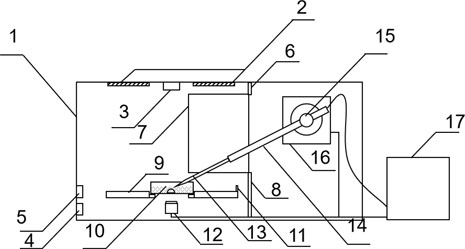
[0021] A detection system for peripheral fluid of embryos, the structure of which is as follows: figure 1 As shown, it includes a housing 1, which is provided with a stage 9, an imaging device 12 and a probe mechanism, and an analysis and control device 17 is arranged outside the casing; the stage 9 is used to place an embryo small dish 10; the probe mechanism is used To position the microprobe 13 to the embryo peripheral fluid in the embryo small dish 10 and detect or obtain its components; the imaging device 12 is used to collect the image information in the embryo small dish 10, locate the embryonic development situation in the field of view and the position of the probe mechanism; analyze and The input end of the control device 17 is connected to the output end of the imaging device 12, and the output end of the analysis and contr...


PUM


Abstract

The invention discloses an embryo peripheral liquid detection system which comprises a shell, and an objective table for placing an embryo small dish, a probe mechanism for detecting embryo peripheral liquid in the embryo small dish and an imaging device for collecting image information of the embryo small dish are arranged in the shell. An analysis and control device for analyzing embryo peripheral liquid and controlling the probe mechanism to work is arranged outside the shell, the input end of the analysis and control device is connected with the output end of the imaging device, and the output end of the analysis and control device is connected with the controlled end of the probe mechanism; the method for detecting the peripheral liquid of the embryo by using the system comprises the following steps: culturing the embryo; embryo observation; assembling a probe; and detecting the embryo peripheral fluid. According to the method, embryo culture can be achieved, metabolites or secretory proteins in peripheral liquid can be detected in real time, the limitation of embryo quality evaluation of traditional embryo morphology is solved, and the reliability of embryo quality evaluation is improved.

Description

technical field [0001] The invention relates to the technical field of embryo detection, in particular to an embryo peripheral fluid detection system. Background technique [0002] Embryo quality is one of the key factors for successful pregnancy. The developmental potential of embryos, that is, the evaluation of embryo quality, directly affects the screening of embryo transfer and pregnancy outcome. [0003] At present, the evaluation of embryo quality is mainly based on the traditional morphological method, that is, according to the law of embryo fertilization and division, the embryo morphology is observed at specific time points, and the embryo quality is evaluated and graded according to the morphology standard, and the selection level is the highest. embryos for transfer. However, this kind of morphological detection has great limitations, relying on the subjective judgment of observers, and there is no accurate and objective evaluation standard; and the so-called mor...

Claims


Application Information

Patent Timeline
Patent Type & Authority Applications(China)
IPC IPC(8): G01N21/84G01N27/26
CPCG01N21/84G01N27/26
Inventor 徐艇郭正飞
Owner 无锡瑞思医疗科技有限公司
Why Eureka
Eureka Blog
Learn More