Eureka AIR delivers breakthrough ideas for toughest innovation challenges, trusted by R&D personnel around the world.

Pressing hemostasis device used after angiocardiography

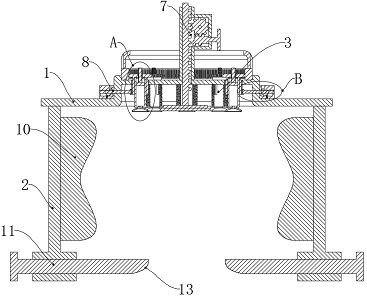
A cardiovascular and air extraction device technology, applied in the field of medical appliances, can solve the problems of secondary wound injury, increase the labor intensity of medical staff, and affect the patient's action, so as to prevent deviation, prevent bleeding, and reduce work intensity.

Inactive Publication Date: 2022-06-07
THE FIRST AFFILIATED HOSPITAL OF ZHENGZHOU UNIV
View PDF0 Cites 0 Cited by
  • Summary
  • Abstract
  • Description
  • Claims
  • Application Information

AI Technical Summary

Problems solved by technology

[0003] After the catheter is withdrawn, it is necessary to pressurize the punctured artery to prevent bleeding. The existing hemostatic device has a relatively simple structure. When the patient's wound bleeds a lot, the hemostatic effect is poor, and the medical staff needs to press for a long time to stop the bleeding, which increases the work of the medical staff. Labor intensity affects the patient's actions at the same time; in addition, some existing hemostatic devices with pressing structures need to lift the patient's limbs in order to install the equipment during actual operation, which will not only cause secondary damage to the patient's wound Injury, and there are still problems that affect the patient's movement, and the existing hemostatic device is not stable in pressing, which is not conducive to the recovery of the wound

Method used


Image

  • Pressing hemostasis device used after angiocardiography
  • Pressing hemostasis device used after angiocardiography
  • Pressing hemostasis device used after angiocardiography

Examples


Embodiment Construction

[0034] In order to further illustrate the various embodiments, the present invention provides accompanying drawings, which are part of the disclosure of the present invention, and are mainly used to illustrate the embodiments, and can be used in conjunction with the relevant descriptions in the specification to explain the operation principles of the embodiments. For these, those of ordinary skill in the art will understand other possible implementations and the advantages of the present invention. Components in the figures are not drawn to scale, and similar component symbols are generally used to represent similar components.

[0035] According to an embodiment of the present invention, a compression hemostasis device after cardiovascular angiography is provided.

[0036] The present invention will now be further described with reference to the accompanying drawings and specific embodiments, such as Figure 1-Figure 12 As shown, the pressure hemostasis device after cardiovas...


PUM


Abstract

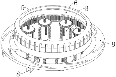
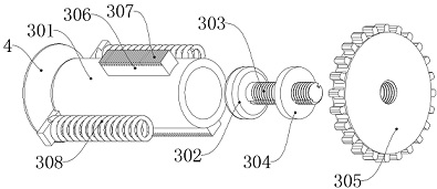
The pressing hemostasis device comprises a supporting plate, and side plates are symmetrically arranged on the two sides of the bottom end of the supporting plate; a plurality of air extracting devices are arranged in the middle of the supporting plate, suckers are arranged at the bottom ends of the air extracting devices, a gear ring is rotationally arranged at the top of the supporting plate, and a pressing device is vertically arranged in the middle of the supporting plate; a plurality of limiting assemblies are arranged in the middle of the supporting plate, turntables are arranged at the top ends of the limiting assemblies, air bags are arranged on the sides, close to each other, of the two side plates, and telescopic plates transversely penetrate through the bottoms of the side plates. The limb of a patient can be wrapped by arranging the supporting plate and the side plates, then the wound of the patient is extruded by using the pressing device, so that the working intensity of medical staff is reduced, the limb of the patient can be wrapped more tightly by using the air bag, the extrusion force of the pressing device is adjustable, the labor-saving effect is achieved, and the medical staff can use the pressing device conveniently. Therefore, the wound bleeding phenomenon of the patient can be effectively prevented.

Description

technical field [0001] The invention relates to the technical field of medical appliances, in particular to a pressure hemostasis device after cardiovascular angiography. Background technique [0002] Cardiovascular angiography is the rapid injection of contrast agents into the heart cavity or blood vessels through the cardiac catheter, which is the development of the heart and blood vessel chambers under X-ray irradiation. The imaging process is photographed. From the imaging results, the blood flow sequence containing the contrast agent and the filling of the cardiac blood vessels can be seen, so as to understand the physiological and anatomical changes of the heart and blood vessels, which is a valuable method for diagnosing cardiovascular diseases. . [0003] After the catheter is withdrawn, it is necessary to pressurize the puncture artery to prevent bleeding. The existing hemostatic device has a relatively simple structure. When the patient bleeds a lot, the hemostati...

Claims


Application Information

Patent Timeline
IPC IPC(8): A61B17/12
CPCA61B17/12A61B2017/12004
Inventor 张春燕王倩谷平平
Owner THE FIRST AFFILIATED HOSPITAL OF ZHENGZHOU UNIV
Why Eureka
Eureka Blog
Learn More