Multipurpose chest adhesion separation device and separation method

A multi-purpose technology for adhesion and separation, applied in the field of medical devices, which can solve the problems of incomplete separation and long separation operation time.

Pending Publication Date: 2022-06-24
SHANDONG PROVINCIAL HOSPITAL AFFILIATED TO SHANDONG FIRST MEDICAL UNIVERSITY
View PDF0 Cites 0 Cited by
  • Summary
  • Abstract
  • Description
  • Claims
  • Application Information

AI Technical Summary

Problems solved by technology

[0003] The purpose of the present invention is to provide a multi-purpose thoracic adhesion separation device and separation method, which are used to solve the problems of long separation operation time and incomplete separation when ordinary biopsy forceps are used to separate adhesions

Method used


Image

  • Multipurpose chest adhesion separation device and separation method
  • Multipurpose chest adhesion separation device and separation method
  • Multipurpose chest adhesion separation device and separation method

Examples


Embodiment 1

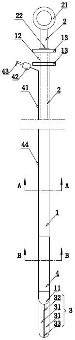
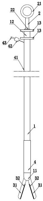
[0042] like Figure 1 to Figure 3 As shown, the cannula 1 is the main body part of the present invention, and the cannula 1 is a hose. One end of the cannula 1 is the insertion part 11, and the other end of the cannula 1 is the operation part 12, such as Figure 8 As shown, the insertion portion 11 of the cannula 1 has a notch 111 , and the biopsy forceps 3 is attached to the insertion portion 11 of the cannula 1 . like Figure 7 As shown, the biopsy forceps 3 includes two forceps handles 31, a blade 32 is fixed on the contact surface of the forceps handles 31, and the blades 32 on the two forceps handles 31 cooperate to cut adhesion or tissue. A plurality of evenly arranged protrusions 33 are also fixed on the contact surface of the forceps handle 31 , and the protrusions on the two forceps handles 31 cooperate to firmly clamp the tissue in the body. And both sides of the blade 32 are provided with protrusions 33, so that after the tissue is clamped by the protrusions on b...

Embodiment 2

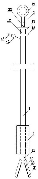
[0050] The difference between the second embodiment and the first embodiment is that in the second embodiment, the biopsy forceps is installed on the slide plate 38, and the slide plate 38 is slidably installed in the cannula 1, and the slide plate 38 is driven in the cannula 1 to move in the cannula 1. expansion unit. Under the action of the telescopic unit, when the sliding plate has the maximum depth in the insertion part, the biopsy forceps is completely retracted into the insertion part, such as Figure 14 As shown, the biopsy forceps are not visually visible at this time. At this time, it is more favorable for the insertion part of the cannula to extend out of the bronchoscope and close to the adhesion site. like Figure 15 , Figure 16 As shown, the forceps handle 31 of the biopsy forceps 3 is rotatably connected with the sliding plate 38, the driving gear 35 and the wire pulley 37 are also rotatably connected with the sliding plate 38, and a telescopic air bag 6 is ...


PUM


Abstract

The invention discloses a multipurpose chest adhesion separation device and a separation method, relates to the technical field of medical instruments, and aims to solve the problems of long separation operation time and incomplete separation when adhesion is separated by using common biopsy forceps. The separating device comprises an intubation tube and an operating rod, the two ends of the intubation tube are an inserting part and an operating part respectively, biopsy forceps are movably installed on the inserting part, one end of the operating rod extends into the operating part of the intubation tube, and a driving unit for driving the biopsy forceps to be closed is arranged between one end of the operating rod and the biopsy forceps. The outer wall of the insertion part is wrapped with a separation air bag, the separation air bag is connected with a connector through a separation air pipe, a plug is detachably installed on the connector, and during use, the plug is detached to inflate the connector so as to inflate the separation air bag. Efficient separation of pleural adhesion is achieved through cooperation of expansion of the separation air bag and shearing of the biopsy forceps. And irregular tumors in the airway can be cut off, and pathological tissues can be obtained.

Description

technical field [0001] The invention relates to the technical field of medical devices, in particular to a multipurpose thoracic cavity adhesion separation device and a separation method. Background technique [0002] The instruments used for the separation of common thoracoscopic adhesions are mostly biopsy forceps used in bronchoscopy biopsy. In the prior art, the opening of the front end of the biopsy forceps is small, which leads to a long time for separation of adhesions, causing the patient to be in pain and suffering for a long time; in addition, using Biopsy forceps are used to cut and separate adhesions, and the workload of doctors is relatively large. When encountering patients with severe adhesions, the effect of adhesion and separation is often poor. For patients with tracheal tumor or granulation, irregular tumor or granulation tissue in the airway was often seen in the past, which made ligation difficult and required electrocution or clamping. On the one hand,...

Claims


Application Information

Patent Timeline
Patent Type & Authority Applications(China)
IPC IPC(8): A61B10/04A61B10/06
CPCA61B10/04A61B10/06
Inventor 吕建利朱文静韩波刘奉琴李经娜
Owner SHANDONG PROVINCIAL HOSPITAL AFFILIATED TO SHANDONG FIRST MEDICAL UNIVERSITY
Why Eureka
Try Eureka