In-vivo implant capable of promoting endothelial cell adhesion and differentiation

A technology of implants and endothelial cells in vivo, applied in the field of implants in vivo, can solve the problems of increased thrombus, achieve the effects of preventing restenosis, better therapeutic effect, and increased implant stability

Active Publication Date: 2022-07-05
开封市卫生学校
View PDF6 Cites 0 Cited by
  • Summary
  • Abstract
  • Description
  • Claims
  • Application Information

AI Technical Summary

Problems solved by technology

That is to say, although the existing drug-coated stents can effectively redu

Method used


Image

  • In-vivo implant capable of promoting endothelial cell adhesion and differentiation
  • In-vivo implant capable of promoting endothelial cell adhesion and differentiation

Examples


Embodiment 1

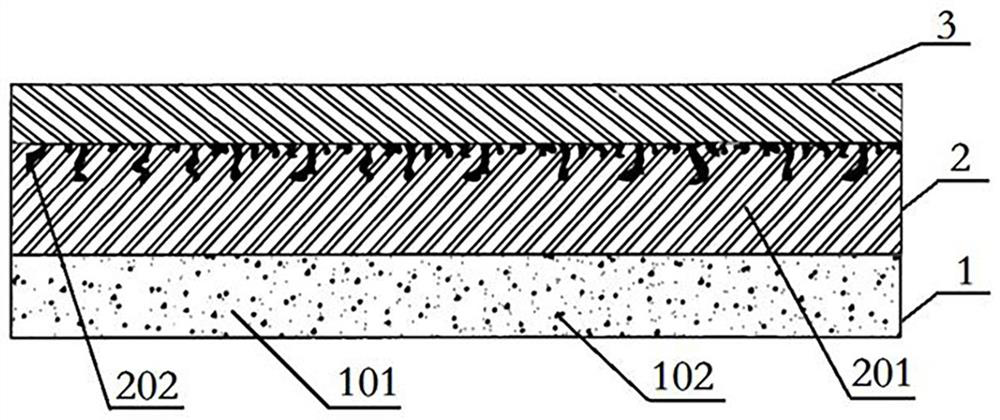
[0023] see attached figure 1 As shown, an in vivo implant that promotes endothelial cell adhesion and differentiation consists of a carrier and a support and includes an inner region 3, an outer region 1, and an intermediate region 2 sandwiched between the inner region 3 and the outer region 1 , the thickness of the middle area 2 is greater than the thickness of the outer area 1, the thickness of the outer area 1 is greater than the thickness of the inner area 3, the middle area 2 and the inner area 3 together constitute the support of the implant, forming the The inner area 3 of the support body is made of degradable material, which can strongly support the implant as a whole. An intermediate area 2 is integrally formed on the outside of the inner area 3, and the intermediate area 2 is made of degradable material. The material 201 is compounded with the first drug 202, and a layer of carrier is sprayed on the periphery of the middle region 2 to form the outer region 1. The ca...

Embodiment 2

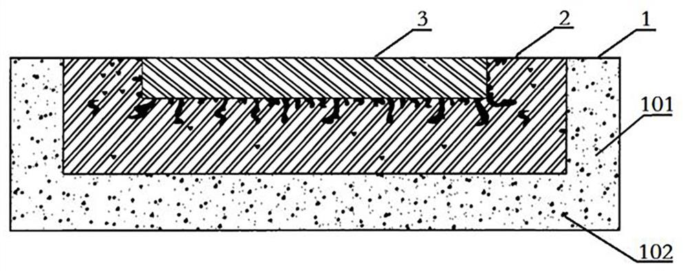
[0034] The structure of this embodiment is basically the same as that of Embodiment 1, the difference is that in this embodiment, as figure 2 As shown, when the implant is a sheet-like structure, its layers can also be folded and wrapped up at the edges after being stacked, that is, the peripheral edges of the outer region 1 are folded upward to wrap the outer edges of the middle region 2, and the middle region The peripheral edges of 2 are folded upward to wrap the outer edges of the inner region 3, and finally an implant with smooth upper and lower surfaces is formed.

[0035] When the degradable material is a polymer of degradable biopolymer materials, degradable magnetic nanoparticles with Fe3O4 as the main material can also be added to the constituent materials of the inner region 3 of the implant, and the degradable magnetic nanoparticles can be made by an external magnetic field. Nanoparticles generate a thermal effect, which in turn can heat up the interior of the imp...


PUM

PropertyMeasurementUnit
Thicknessaaaaaaaaaa
Thicknessaaaaaaaaaa
Thicknessaaaaaaaaaa

Abstract

The invention discloses an in-vivo implant capable of promoting adhesion and differentiation of endothelial cells, an internal area of the implant is made of a degradable material and plays a role in throughout and always supporting the implant, a middle area is formed by compounding the degradable material and a first drug, and an external area is formed by compounding an organic polymer material and a second drug and plays a role in throughout and always supporting the implant. The first medicine is an anti-restenosis medicine for inhibiting smooth muscle cell proliferation, the second medicine is a specific endothelial progenitor cell antibody capturing medicine, and the proportion of the second medicine in an external area is gradually changed, so that the adhesion and differentiation capabilities of endothelial cells around a tissue injury part are improved, and the tissue injury effect is improved. And meanwhile, after the expected repairing purpose is achieved, the slow release of the first medicine is utilized to inhibit the cell proliferation so as to prevent the occurrence of restenosis. The variable stacking form can adapt to the structural characteristics of the human body, the requirements of different patients and different implantation positions are met, the operation is safer and more reliable, and the postoperative curative effect is better.

Description

technical field [0001] The present invention relates to a medical implant, especially an implant in vivo which can promote the adhesion and differentiation of endothelial cells. Background technique [0002] Promoting cell adhesion and differentiation plays a prominent role in the biomedical field, especially in the field of in vivo implants. Simple pure metal stents lacking cell adhesion and differentiation functions gradually lose their superiority. Drug-coated stents have become the current The internationally recognized clinical preferred method, and the commonly used drug-eluting stents mainly include non-degradable and degradable carrier drug-eluting stents. Drug-eluting stents bring hope for reducing or eliminating restenosis. However, no matter what kind of stent is implanted, it will cause damage to endothelial cells and smooth muscle cells in the lumen wall, which is likely to cause thrombosis and inflammatory reactions. The release of cell growth factors accompan...

Claims


Application Information

Patent Timeline
IPC IPC(8): A61L31/12A61L31/06A61L31/02A61L31/14A61L31/16A61L27/40A61L27/18A61L27/02A61L27/04A61L27/50A61L27/54A61L27/58
CPCA61L31/06A61L31/028A61L31/022A61L31/14A61L31/148A61L31/16A61L27/18A61L27/025A61L27/047A61L27/50A61L27/507A61L27/54A61L27/58A61L2300/412A61L2300/42A61L2300/416A61L2300/256A61L2300/608A61L2430/02A61L2430/38
Inventor 贺晓静轩文娟曹馨月曹守威
Owner 开封市卫生学校
Why Eureka
Try Eureka