
Method for compressing human body pipeline tissue three-dimensional model

A technology of 3D model and compression method, which is applied in the medical field, can solve problems affecting the real effect of 3D rendering, large 3D model file size, and spike phenomenon, and achieve the effects of reduced network transmission performance, good surgical planning, and reduced quantity

Pending Publication Date: 2022-07-05
诺信医学科技(山东)有限公司
View PDF0 Cites 0 Cited by
  • Summary
  • Abstract
  • Description
  • Claims
  • Application Information

AI Technical Summary

Problems solved by technology

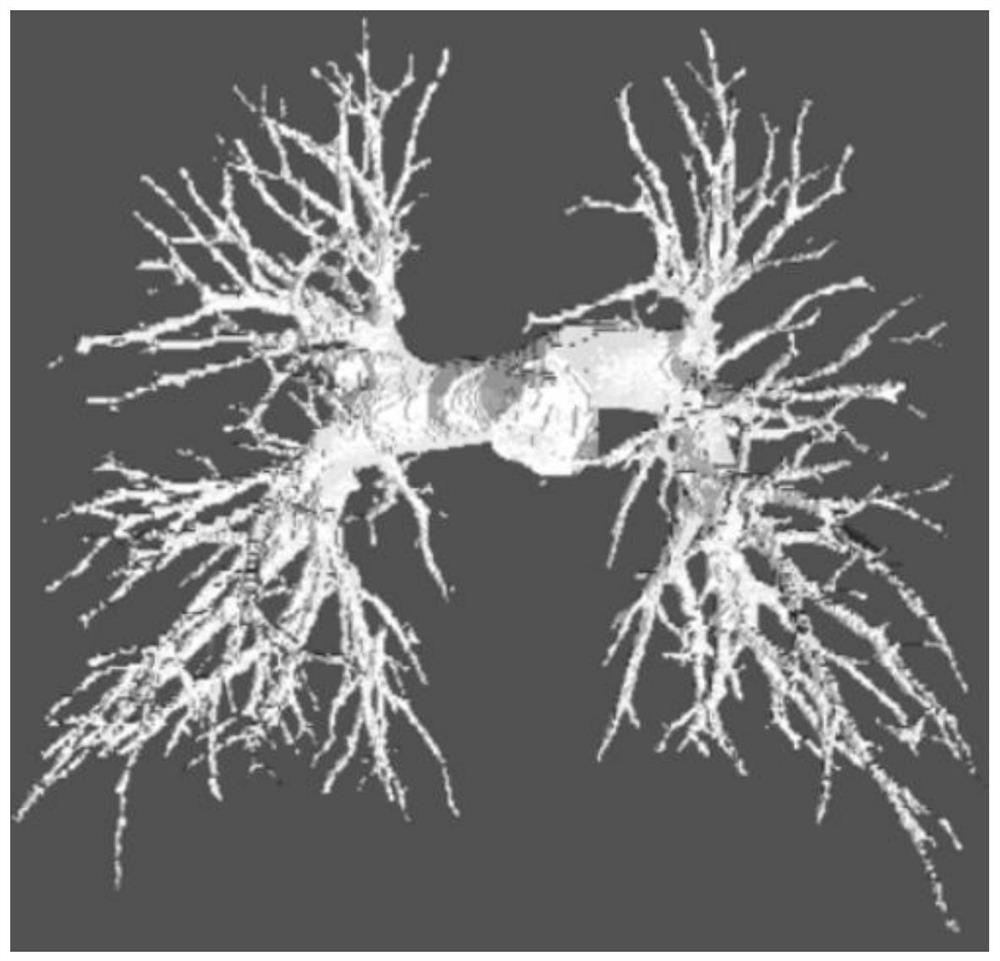
[0004] However, human organs, especially blood vessels, bronchi and other pipelines, have complex structures and are distributed in dendrites. Due to the large average curvature of the surface, the 3D model needs more and finer patches to achieve smooth surface fitting; this makes the 3D model The file size is large. For example, the unprocessed 3D model of the pulmonary artery often reaches several 10M bytes, and it can reach hundreds of Mbytes after smoothing algorithm processing; large-size 3D files are essential for database storage, 3D interactive algorithm design and mobile medical The transmission of the application generates a lot of pressure; on the other hand, if the original 3D reconstruction model is directly smoothed, the model may be shrunk, and spikes will appear at the end of the pipeline tissue, which will affect the real effect of 3D rendering

Method used


Image

  • Method for compressing human body pipeline tissue three-dimensional model
  • Method for compressing human body pipeline tissue three-dimensional model
  • Method for compressing human body pipeline tissue three-dimensional model

Examples


Embodiment 1

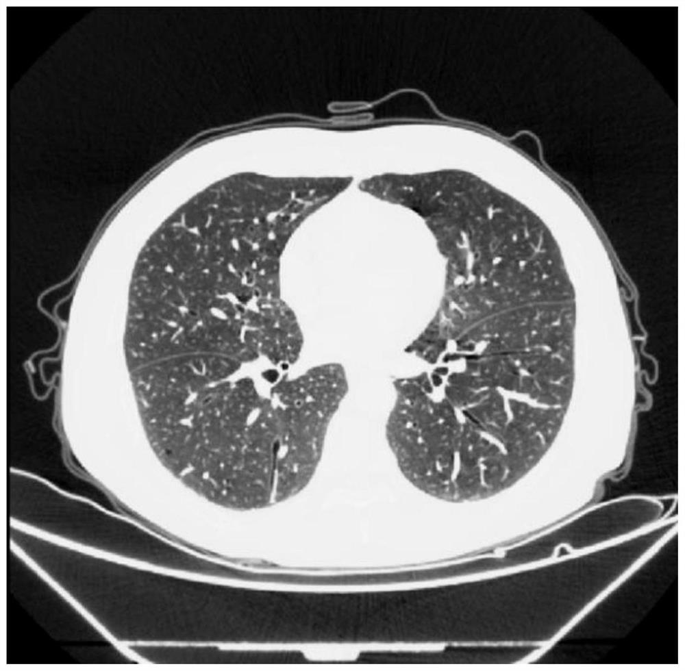
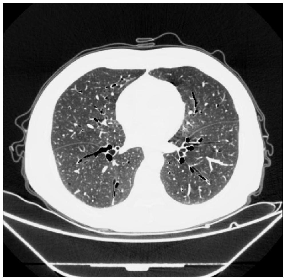
[0032] like Figure 1-5 As shown, a method for compressing a three-dimensional model of a human body pipeline tissue includes the following specific steps:

[0033] Step 1: Obtain the preoperative CT images of the patient, perform homogeneity resampling, and normalize CT values ​​for preprocessing;

[0034] Step 2: Input the CT sequence into the pre-trained image segmentation algorithm to extract the target tissue; segment it into a target tissue mask image sequence, that is, the voxels belonging to the target tissue in the original medical image are marked in white (8-bit code). , grayscale value = 255);

[0035] Step 3: Perform connectivity denoising processing. According to the connectivity of voxels 6, the first N largest connected domains are taken, and other smaller connected domains are removed as segmentation noise; N is set as 2;

[0036] Step 4: Perform Gaussian smoothing on the segmentation mask to obtain grayscale mask data whose grayscale transitions from 255 t...

Embodiment 2

[0048] like Figure 1-5 As shown, a method for compressing a three-dimensional model of a human body pipeline tissue includes the following specific steps:

[0049] Step 1: Obtain the preoperative CT images of the patient, perform homogeneity resampling, and normalize CT values ​​for preprocessing;

[0050] Step 2: Input the CT sequence into the pre-trained image segmentation algorithm to extract the target tissue; segment it into a target tissue mask image sequence, that is, the voxels belonging to the target tissue in the original medical image are marked in white (8-bit code). , grayscale value = 255);

[0051] Step 3: Perform connectivity de-noising processing. According to the connectivity of the voxels, the 8 connected domains are selected, and the first N largest connected domains are taken, and the other smaller connected domains are removed as segmentation noise; N is set as 3;

[0052] Step 4: Perform Gaussian smoothing on the segmentation mask to obtain grayscale...

Embodiment 3

[0064] like Figure 1-5 As shown, a method for compressing a three-dimensional model of a human body pipeline tissue includes the following specific steps:

[0065] Step 1: Obtain the preoperative CT images of the patient, perform homogeneity resampling, and normalize CT values ​​for preprocessing;

[0066] Step 2: Input the CT sequence into the pre-trained image segmentation algorithm to extract the target tissue; segment it into a target tissue mask image sequence, that is, the voxels belonging to the target tissue in the original medical image are marked in white (8-bit code). , grayscale value = 255);

[0067] Step 3: Perform connectivity de-noising processing. According to the connectivity of the voxels, the 8 connected domains are selected, and the first N largest connected domains are taken, and the other smaller connected domains are removed as segmentation noise; N is set as 4;

[0068] Step 4: Perform Gaussian smoothing on the segmentation mask to obtain grayscale...


PUM


Abstract

The invention discloses a human body pipeline tissue three-dimensional model compression method, which comprises the following steps: acquiring a preoperative detection CT image of a patient, and carrying out isotropic resampling and CT value normalization preprocessing; inputting the CT sequence into a pre-trained image segmentation algorithm, and extracting a target tissue; segmenting into a target tissue mask image sequence, namely marking voxels belonging to the target tissue in the original medical image as white (8-bit coding, and gray scale value = 255); performing communication de-noising processing, according to voxel connectivity 6 communication or 8 communication, taking the first N maximum connected domains, and taking other smaller connected domains as segmentation noise to be removed; n is set to be 2-4 according to different tissues and different image quality characteristics. According to the method, the number of surface patches can be greatly reduced while the rendering smooth effect of the three-dimensional model is not seriously reduced, the size of the three-dimensional model is reduced, and the use efficiency is improved.

Description

technical field [0001] The invention belongs to the field of medical technology, and in particular relates to a method for compressing a three-dimensional model of a human body duct tissue. Background technique [0002] Three-dimensional reconstruction of medical images is a process of generating surface patch fitting models of human organs through organ segmentation and three-dimensional modeling based on human tomography (CT or MRI). The polygonal patch is a digital model of the surface shape of the human body's three-dimensional organs; it can be drawn on a computer screen by a three-dimensional rendering method to present a three-dimensional image of human organs and realize the observation effect of virtual reality; the patch is the basic unit of the three-dimensional model of the organ , many tiny patches are connected to each other to fit the surface of 3D organs; patch smoothing is a processing algorithm for 3D reconstruction model surface patches, which can make the...

Claims


Application Information

Patent Timeline
Patent Type & Authority Applications(China)
IPC IPC(8): G06T19/20G06T17/00G06T15/00G06T7/10G06T5/00
CPCG06T19/20G06T17/00G06T15/005G06T7/10G06T2207/10081G06T5/70
Inventor 田光野徐栗满海堂巩道友
Owner 诺信医学科技(山东)有限公司
Learn More