Esophageal-gastric junction tumor image classification method, system and device and storage medium
A gastric junction and junction technology are applied in the field of image classification methods, equipment and storage media, and systems for tumors in the esophagogastric junction, which can solve problems such as limited research in identification, reduce pain and economic burden, and stabilize evaluation effects. Easy-to-operate effects
- Summary
- Abstract
- Description
- Claims
- Application Information
AI Technical Summary
Problems solved by technology
Method used
Image
Examples
Embodiment 1
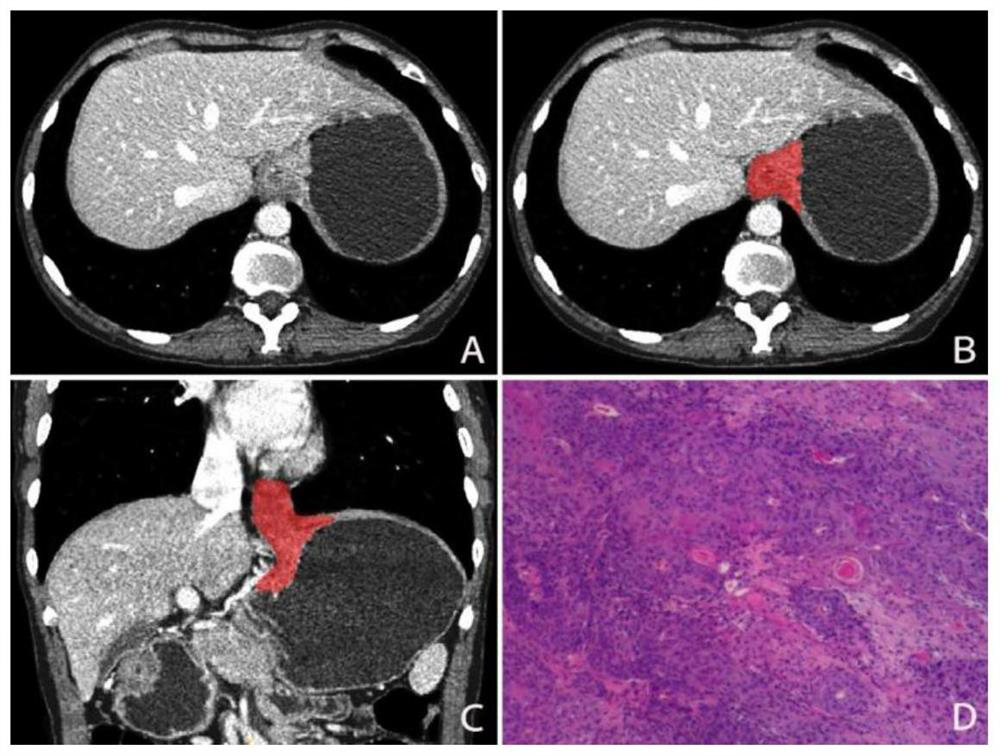
[0149] A method for classifying images of esophagogastric junction tumors, comprising the following steps:
[0150] S1. Process the enhanced CT image of the tumor at the esophagogastric junction of the subject to obtain a three-dimensional ROI area image of the tumor medical image lesion; the enhanced CT image is an arterial phase enhanced CT image, and the three-dimensional ROI area image is an arterial phase image 3D ROI area image;
[0151] S2, extracting the radiomics feature in the three-dimensional ROI area image;
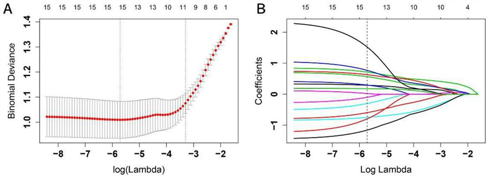
[0152] S3, the value of the radiomics feature extracted in step S2 is input into the score prediction model, and the image score of the three-dimensional ROI region image is obtained by calculation;
[0153] S4. Perform qualitative analysis on the image scores obtained in step S3 to predict the image type of the tumor medical image.
[0154]In step S1, the tumor medical image of the esophagogastric junction of the subject is processed, and the specific oper...
Embodiment 2
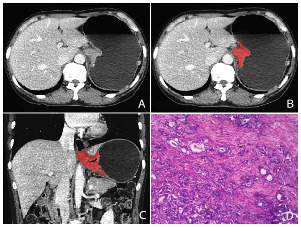
[0198] A method for classifying images of esophagogastric junction tumors, comprising the following steps:
[0199] S1. Process the enhanced CT image of the tumor at the esophagogastric junction of the subject to obtain a three-dimensional ROI area image of the tumor medical image lesion; the enhanced CT image is a venous phase enhanced CT image, and the three-dimensional ROI area image is a venous phase image 3D ROI area image;
[0200] S2, extracting the radiomics feature in the three-dimensional ROI area image;
[0201] S3, the value of the radiomics feature extracted in step S2 is input into the score prediction model, and the image score of the three-dimensional ROI region image is obtained by calculation;
[0202] S4. Perform qualitative analysis on the image scores obtained in step S3 to predict the image type of the tumor medical image.
[0203] In step S1, the tumor medical image of the esophagogastric junction of the subject is processed, and the specific operation...
Embodiment 3
[0236] A method for classifying images of esophagogastric junction tumors, comprising the following steps:
[0237] S1. Process the enhanced CT image of the tumor at the esophagogastric junction of the subject to obtain a three-dimensional ROI image of the tumor medical image lesion; the enhanced CT image includes an arterial phase enhanced CT image and a venous phase enhanced CT image, and the three-dimensional The ROI area images include three-dimensional ROI area images in the arterial phase and three-dimensional ROI area images in the venous phase. The enhanced CT images in the arterial phase of the subjects' esophagogastric junction tumors are processed to obtain the three-dimensional ROI area images in the arterial phase. The venous phase enhanced CT images of the junction tumor were processed to obtain a three-dimensional ROI image in the venous phase;
[0238] S2, extracting the radiomic features in the three-dimensional ROI area image of the arterial phase and the thr...
PUM
 Abstract
Description
Claims
Application Information
 - R&D
- Intellectual Property
- Life Sciences
- Materials
- Tech Scout
- Unparalleled Data Quality
- Higher Quality Content
- 60% Fewer Hallucinations
Browse by: Latest US Patents, China's latest patents, Technical Efficacy Thesaurus, Application Domain, Technology Topic, Popular Technical Reports.
© 2025 PatSnap. All rights reserved.Legal|Privacy policy|Modern Slavery Act Transparency Statement|Sitemap|About US| Contact US: help@patsnap.com


