Ear-nose-throat visual inspection equipment

An inspection equipment and ENT technology, applied in the field of medical devices, can solve problems such as inability to inspect the angle and position adjustment, inability to visually inspect the affected area, and affect the inspection of medical staff, so as to improve inspection efficiency, flexible use, and good comfort Effect

Pending Publication Date: 2022-08-05
XIEHE HOSPITAL ATTACHED TO TONGJI MEDICAL COLLEGE HUAZHONG SCI & TECH UNIV
View PDF1 Cites 0 Cited by
  • Summary
  • Abstract
  • Description
  • Claims
  • Application Information

AI Technical Summary

Problems solved by technology

[0003] A kind of otolaryngology examiner disclosed by the invention of authorization announcement number as CN106215311B, comprises a first mounting plate, and said first mounting plate is provided with mounting hole, and a ring-shaped lamp tube is installed around the mounting hole, and the mounting hole There is a circle of reflection plate installed on the inner side, and a circle of elastic fixed ring is installed in the installation hole, and a transparent sleeve that is in sealing contact with the fixed ring is penetrated into the installation hole. There is an inspection slot at the front end, and a strip slot is connected to the rear end of the inspection slot; the transparent sleeve is equipped with an applicator, but it cannot be fixed in the ear canal, nasal cavity and oral cavity, and the fixation stability is not good, which affects medical care Personnel inspection, at the same time, it cannot adjust the inspection angle and position, the inspection accuracy is not high, and it is impossible to visually inspect the patient's affected area, and the inspection effect is not good. Therefore, we propose a visual inspection equipment for ENT

Method used


Image

  • Ear-nose-throat visual inspection equipment
  • Ear-nose-throat visual inspection equipment
  • Ear-nose-throat visual inspection equipment

Examples


Embodiment 1

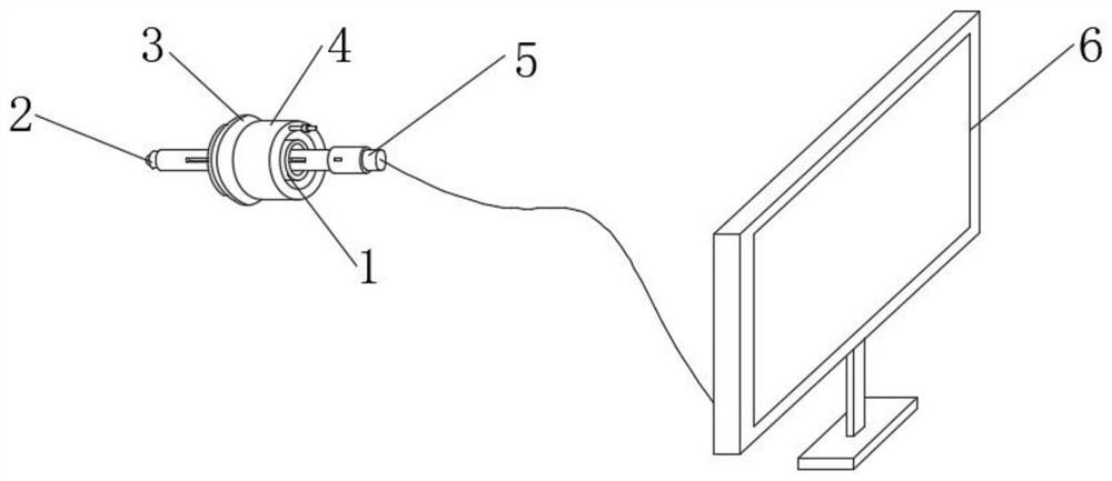
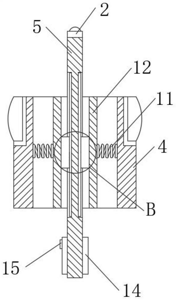
[0033] see Figure 1-Figure 7 , the present invention provides a technical solution: an ENT visual inspection device, comprising an installation sleeve 4, an adjustment component 1 for adjusting the position and angle of the inspection rod 5 is arranged in the installation sleeve 4, and a microporous probe 2 is arranged at one end of the inspection rod 5 , the microporous probe 2 is connected with the display terminal 6 through a connecting line, and a detachable fixing assembly 3 is arranged on the outer surface of the installation sleeve 4. The detachable fixing assembly 3 is used for the installation sleeve 4 to be fixed in the patient's ear canal, nasal cavity or oral cavity.

[0034] The adjustment assembly 1 includes a support sleeve 12, the support sleeve 12 is arranged in the installation sleeve 4, and the outer surface of the installation sleeve 4 is provided with a return spring 11 in an annular and equidistant manner. The metal slider 13 is arranged symmetrically on...

Embodiment 2

[0046] see Figure 8 , the fixed structure is an upper support block 32 and a lower support block 35, the upper support block 32 is arranged at one end of the upper pole 31, the lower support block 35 is arranged at one end of the lower pole 34, and the upper pole 31 and the lower pole 34 are symmetrically arranged on the bearing On both sides of the outer surface of the ring 302 , upper tooth positioning grooves 33 are arranged on the upper side of the upper support block 32 , and lower tooth positioning grooves 36 are arranged on the lower side of the lower support block 35 .

[0047] Working principle, when the throat of the patient needs to be checked, the carrier ring 302 with the upper support block 32 and the lower support block 35 is selected, and then the positioning block 303 on the carrier ring 302 is snapped into the guide groove 304. When one end of the clamping block 303 moves to the position of the positioning groove 308, the bearing ring 302 is attached to the ...


PUM


Abstract

The invention discloses ear-nose-throat visual inspection equipment which comprises a mounting sleeve, an adjusting assembly used for adjusting the position and angle of an inspection rod is arranged in the mounting sleeve, a micropore probe is arranged at one end of the inspection rod and connected with a display terminal through a connecting line, a detachable fixing assembly is arranged on the outer surface of the mounting sleeve, and the detachable fixing assembly is connected with the display terminal through a connecting line. The detachable fixing assembly is used for fixing the mounting sleeve in the ear canal, the nasal cavity or the oral cavity of the patient, the throat, the nasal cavity and the ear canal of the patient can be visually examined, the examination efficiency is improved, meanwhile, the examination position and the examination angle can be adjusted, the examination omnibearing performance is guaranteed, and the examination efficiency is improved. The device can be rapidly fixed in the oral cavity, the nasal cavity and the ear canal of a patient, is good in fixing stability, convenient to operate, good in use comfort degree and more flexible to use, and meets the requirement for examination of patients at different parts.

Description

technical field [0001] The invention belongs to the technical field of medical devices, and in particular relates to a visual inspection device for ear, nose and throat. Background technique [0002] At present, when examining the patient's ear cavity and nasal cavity, medical staff use a flashlight in their hand to illuminate, and only observe directly through the eyes; it is more troublesome to examine the oral cavity and throat, usually holding a tongue depressor in one hand and pressing the patient down. Tongue, hold a flashlight in the other hand into the patient's mouth and throat for observation, or use the reflector worn on the head to reflect the light into the patient's mouth or throat. [0003] For example, an ENT examiner disclosed by the invention with the authorization announcement number of CN106215311B includes a first mounting plate, the first mounting plate is provided with a mounting hole, and a ring-shaped lamp tube is mounted around the mounting hole, an...

Claims


Application Information

Patent Timeline
Patent Type & Authority Applications(China)
IPC IPC(8): A61B1/227A61B1/233A61B1/267A61B1/24A61B1/04A61B1/00
CPCA61B1/227A61B1/233A61B1/267A61B1/24A61B1/04A61B1/00131A61B1/00147
Inventor 鲁才红王峥嵘王勤林婉婷胡梦云熊沙沙
Owner XIEHE HOSPITAL ATTACHED TO TONGJI MEDICAL COLLEGE HUAZHONG SCI & TECH UNIV
Why Eureka
Try Eureka