Method for segmenting anatomical structures from 3d image data by using topological information

A three-dimensional image, anatomical structure technology, applied in image data processing, image data processing, image analysis and other directions, can solve problems such as inability to reach blood vessels

Inactive Publication Date: 2006-03-15
SIEMENS AG
View PDF0 Cites 12 Cited by
  • Summary
  • Abstract
  • Description
  • Claims
  • Application Information

AI Technical Summary

Problems solved by technology

[0005] Known "region growing" techniques work satisfactorily in many cases, but do not reach all blood vessels visible to the viewer in the display of the 3D image photograph
In proper 3D visualization techniques, the human eye is still able to identify the smallest blood vessels as part of the

Method used


Image

  • Method for segmenting anatomical structures from 3d image data by using topological information
  • Method for segmenting anatomical structures from 3d image data by using topological information
  • Method for segmenting anatomical structures from 3d image data by using topological information

Examples


Embodiment Construction

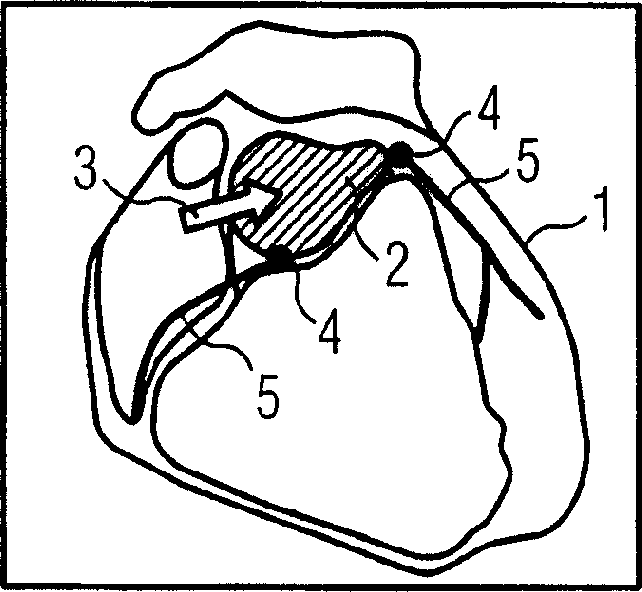
[0020] In this example, the individual steps for segmenting a coronary vessel tree from three-dimensional image data recorded with CTA techniques are described. The figures here show different views of the heart with the atria and blood vessels contained therein, which are only shown schematically due to the lack of visualization of the image itself. here figure 1 A view of the entire heart 1 is shown, a section through the aorta 2 being visible at the top. In a first step, a starting point is set, eg interactively with the mouse cursor 3 , by clicking on the aorta 2 on the screen where the image is displayed. Starting from this starting point, two bifurcation points 4 are automatically detected at which the aorta 2 diverges into coronary vessel structures 5 . Mark the bifurcation point 4.

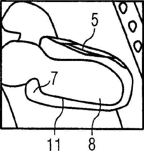
[0021] In the next step, seen with the help of parts of FIGS. 2A, 2B, 2C, the atrium 11, that is, the left and right atrium and the ventricle, is segmented and anatomical landmarks, in ...


PUM


Abstract

A method is disclosed for segmenting anatomical structures, in particular the coronary vessel tree, from 3D image data. In the method, a starting point is initially set in the 3D image data, and at least one known anatomically significant point and/or at least one known anatomically significant surface are/is identified in the 3D image data. Subsequently, proceeding from the starting point the structure is subsequently segmented pixel by pixel with the aid of a multiplicity of segmentation steps in such a way that an instantaneous distance is determined automatically relative to the anatomically significant point and/or to the anatomically significant surface in each segmentation step. Further, segmentation parameters and/or a selection of adjacent pixels for continuing the segmentation are/is established as a function of the distance, taking account of a model topology. The method enables an accurate and reliable segmentation of the anatomical structure.

Description

technical field [0001] The invention relates to a method for segmenting anatomical structures, in particular coronary vascular trees, from three-dimensional image data, for example generated in CT angiography (CTA). [0002] The method according to the invention is firstly used in the field of computed tomography when recording vascular structures. Background technique [0003] Compared with other imaging techniques such as Magnetic Resonance Tomography (MR), PET (Positron Emission Tomography), SPECT (Single Photon Emission Computed Tomography) or 3D ultrasound techniques, the great advantage of CT angiography is e.g. The entire vascular tree of the heart can be documented with only one CT scan in the absence of drugs. The three-dimensional image data obtained here can be visualized using different techniques. [0004] For quantitative analysis, especially for measuring stenosis or plaque deposition, it is necessary to segment the corresponding regions of the vascular stru...

Claims


Application Information

Patent Timeline
IPC IPC(8): A61B6/03G06T1/00G06T15/00A61B5/00A61B5/055A61B8/00G06T7/60
CPCG06T7/0081G06T2207/20101G06T2207/10081G06T2207/30101G06T7/11
Inventor 丹尼尔·林克迈克尔·肖伊林
Owner SIEMENS AG
Why Eureka
Try Eureka