A method and kit for detecting the early onset of renal tubular cell injury

A technology of renal tubular cells and kits, which can be used in biological testing, material testing, etc., and can solve problems such as Cyr61 detection problems.

Inactive Publication Date: 2006-06-21
CHILDRENS HOSPITAL MEDICAL CENT CINCINNATI +1
View PDF5 Cites 50 Cited by
  • Summary
  • Abstract
  • Description
  • Claims
  • Application Information

AI Technical Summary

Problems solved by technology

Therefore, detection of Cyr61 in urine is problematic in te...

Method used


Image

  • A method and kit for detecting the early onset of renal tubular cell injury
  • A method and kit for detecting the early onset of renal tubular cell injury
  • A method and kit for detecting the early onset of renal tubular cell injury

Examples


Embodiment 1

[0099] NGAL is a small protease-resistant, secreted polypeptide that can be detected in urine. Significant upregulation of NGAL mRNA and protein levels has been shown early after renal ischemia in mice. NGAL protein expression was mainly detected in the proximal tubule cells, which resembled a secreted protein with a punctate distribution in the cytoplasm. Indeed, in mouse and rat models of ARF, NGAL is readily and rapidly detectable in urine (in the first urination) after ischemic injury, when no renal leukocytes are observed penetration. The origin of NGAL from tubular cells was further confirmed in cultured human proximal tubular cells subjected to in vitro ischemic injury, where NGAL mRNA was significantly and immediately Inside, NGAL protein was readily detectable in the culture medium. Our results illustrate that NGAL may represent a new early urinary biomarker of ischemic kidney injury.

[0100] Identification of novel genes upregulated early after renal ischemia-re...

Embodiment 2

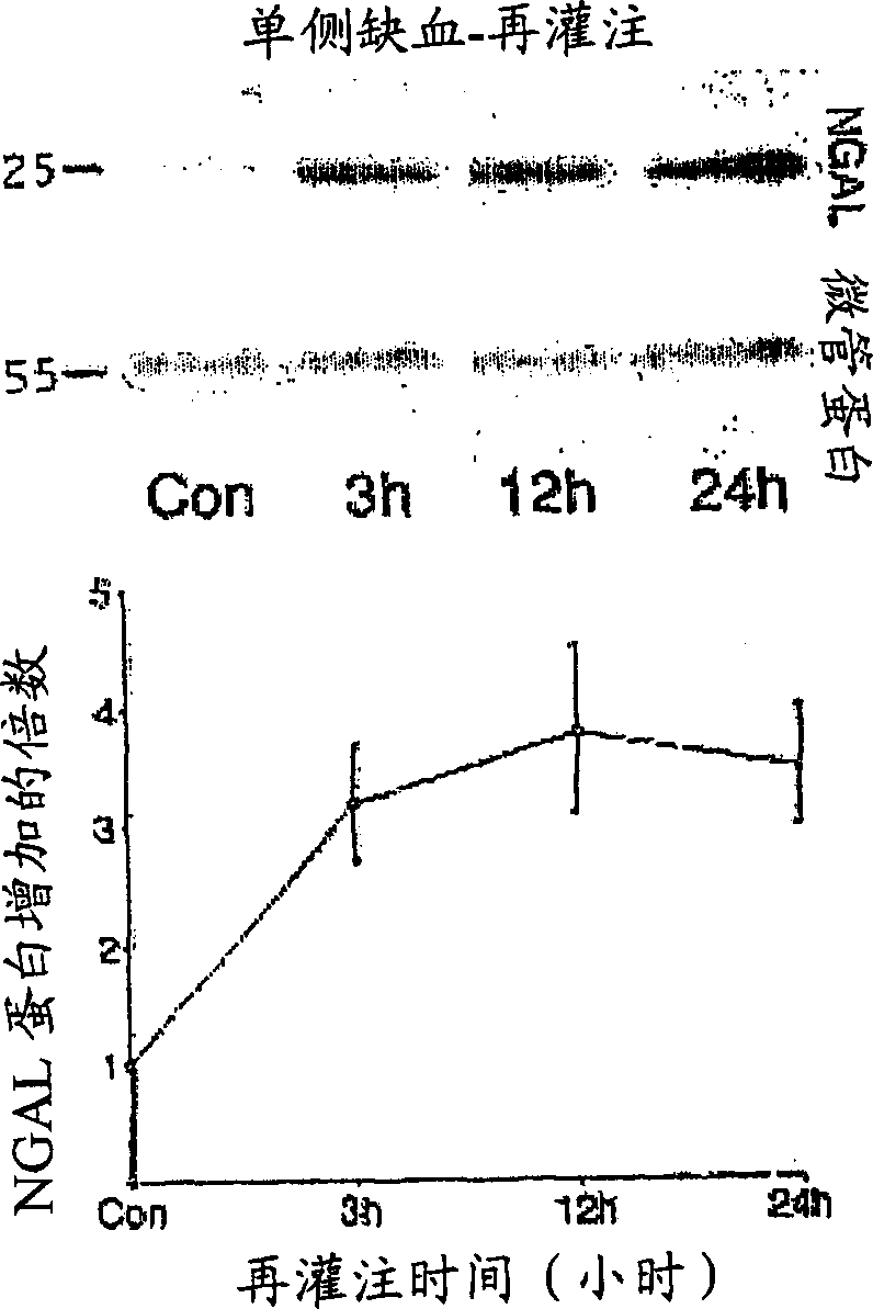
[0114] NGAL protein was readily detected in urine immediately after induction of ARF in rats:

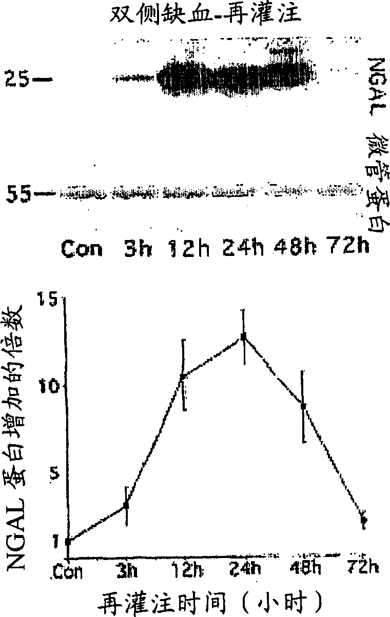
[0115] Because of the debate regarding species differences in the response to renal artery occlusion, we below examined the behavior of NGAL in a different animal model, an established rat model of renal ischemia-reperfusion injury. Urinary creatinine concentrations were used to make loading consistent, and NGAL was absent in urine prior to renal ischemia in rats. In marked contrast, NGAL showed 25 kDa immunoreactivity within 3 hours of injury (in the first post-ischemic urine effusion), as shown in Figure 6 shown. In comparison, serum creatinine in this model of ischemic injury was elevated only after 24 hours of reperfusion (not shown). Again, NGAL was detectable in as little as 1 [mu]l of untreated urine and persisted throughout the period examined (24 hours of reperfusion).

Embodiment 3

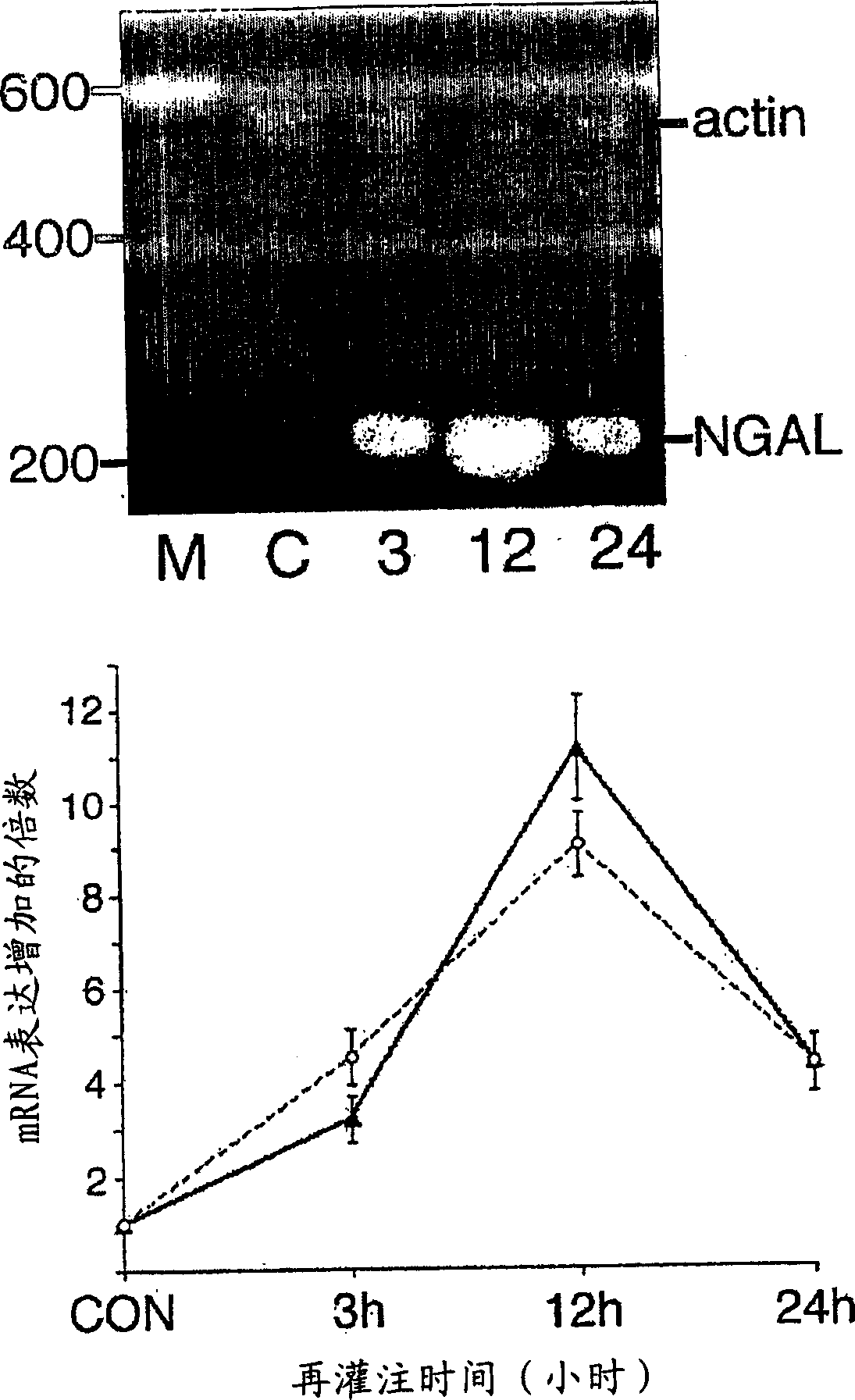
[0117] NGAL mRNA is induced in cultured human proximal tubule cells after early minimal ischemia:

[0118]To confirm the source of NGAL from ischemic proximal tubular cells, we modified a previously described procedure for in vitro ischemia induced by ATP depletion in cultured human proximal tubular cells (RPTEC). Incubation in 1 μM antimycin produced a mild partial ATP depletion, which became about 83±3% of the control within 1 hour and decreased more slowly to about 75±3% of the control by 6 hours (from four Mean + / - SD of experiments). The morphological consequences of this mild ATP depletion were not discernible. NGAL mRNA is barely detectable in resting cells. However, after partial ATP depletion, a rapid duration-dependent induction of NGAL mRNA was clearly visible by RT-PCR, as shown in Figure 7 shown.

[0119] NGAL protein was readily detected in culture medium following early ischemia in vitro:

[0120] We next examined NGAL protein expression in RPTEC cells and...


PUM


Abstract

An early-onset method and kit for detecting renal tubular cell injury, using NGAL as an early urinary biomarker. NGAL is a small secreted polypeptide that is protease resistant and thus readily detected in urine following tubular cell injury. NGAL protein expression was mainly detected in the proximal tubule cells, which resembled a secreted protein with a punctate distribution in the cytoplasm. Apparent NGAL in urine correlates with the amount and duration of renal ischemia and nephrotoxicemia and is a diagnostic feature of tubular cell injury and renal failure. NGAL detection is also a useful marker for monitoring nephrotoxic side effects of drugs or other therapeutic agents.

Description

Background technique [0001] Acute renal failure (ARF) secondary to renal tubular cell injury, including ischemic or nephrotoxic injury, remains a challenge in clinical medicine and nephrology ( nephrology) with persistently high mortality and morbidity. Decades of previous studies have illustrated the role of persistent vasoconstriction, tubular obstruction, cellular structural and metabolic changes, and inflammatory responses in the pathogenesis of ARF. While these studies have shown possible therapeutic approaches in animal models, efforts to translate them into humans have yielded disappointing results. Reasons for this may include the multifaceted renal response to ischemic injury and nephrotoxins, and the lack of early biomarkers of ARF, resulting in delayed initiation of treatment. [0002] When the patient's serum creatinine (creatinine) value or: (1) when the baseline serum creatinine level is less than 2.0mg / dL, an increase of at least 0.5mg / dL; (2) when the baselin...

Claims


Application Information

Patent Timeline
IPC IPC(8): G01N33/53
Inventor 普拉塞德·德瓦拉简乔纳森·M·巴拉施
Owner CHILDRENS HOSPITAL MEDICAL CENT CINCINNATI
Why Eureka
Try Eureka