Percutaneous access pathway system and method

a percutaneous access and pathway technology, applied in the field of medical devices, can solve the problems of long practice, damage to the underlying structure, and risk of laceration of the underlying organ, and achieve the effects of reducing friction and/or the chance of tissue pinching, reducing user error, and increasing and/or reducing the cross-sectional area of the catheter

Active Publication Date: 2018-08-14
CRITICAL INNOVATIONS
View PDF91 Cites 12 Cited by
  • Summary
  • Abstract
  • Description
  • Claims
  • Application Information

AI Technical Summary

Benefits of technology

[0026]In some embodiments, the percutaneous access pathway is established via the use of a probe and needle mechanism that automatically stops the advance of the device upon insertion into a body cavity or space (e.g. pleural space), such as also described in U.S. Publication No. 2014 / 0046303. This minimizes user error and allows for the device to be used on differently sized patients (e.g. with different sized chest walls). In some embodiments, one device fits all patients. In others, part (e.g. changeable access pathways of various sizes on a reusable or standard insertion device) or all of the device is differently sized for different subgroups so that the appropriately sized device can be chosen for different subgroups based on, for example, weight, age, gender, length, pre-determined size categories (e.g. Broselow scale), and / or other indicators. These differently sized components may come together in a kit, with means for determining proper sizing. Under different embodiments, the insertion device is disposable or non-disposable and different catheters and / or ports may fit onto one insertion device or have their own sized insertion devices.
[0027]Under various embodiments, the percutaneous access pathway itself is made of different materials and methods. Under some embodiments, the percutaneous access pathway is a space (potential or maintained) through body tissue; is a deformable or non-deformable catheter; is expandable and / or deflateable plastic or metal (e.g. stent, mesh, rolled material, reinforced wires); and / or is an expandable and / or deflateable catheter filled with gas (e.g. air) or fluid (e.g. normal saline). Additionally, under some embodiments the catheter is covered partially or fully with additional material(s), flexible in some cases, that can provide additional benefits when in contact with the body tissue. Examples include means to increase and / or decrease the cross-sectional area of the catheter; to reduce friction and / or the chances of tissue being pinched in the underlying catheter or mechanism; to decrease the chances of infection (e.g. antimicrobial properties); and to have drug-releasing properties (e.g. anesthetic or other anti-pain medications).
[0029]In many embodiments, the access pathway is made of a catheter and access pathway port. In some embodiments, the access pathway port allows entrance to the catheter to be reversibly or irreversibly sealed, thus preventing air and / or infection from entering the body cavity. In some embodiments, the connection of an attachment device to the access pathway port causes the access pathway port to open, thus allowing part or all of the attachment device (e.g. chest tube, surgical equipment, endoscopy camera, video-assisted thoracoscopic surgery device) to enter the body through the catheter, but not allowing air or infectious material to enter when an attachment device is not attached. In some embodiments the access pathway port and / or catheter has one or more check valves, which allow air to release from the body but not to enter the body. Under some embodiments, the access pathway port connection uses a quick connect type mechanism, to expedite attachment and simplify the procedure.

Problems solved by technology

The chest tube may be inserted by pushing it through the chest wall over a sharp trocar, although this risks lacerating the underlying organs with the sharp trocar.
This has less chance of damaging vital structures, but is lengthy in practice.
All current methods share some drawbacks including the aforementioned risks of damaging underlying structures, as well as the need to perform tube thoracostomy under a relatively large sterile field due to chest tube length.
This makes it difficult to maintain sterility outside of the operating room, for example in the emergency department, in the out-of-hospital arena, or on the battlefield.
Additionally, the portion of the chest tube outside the body immediately becomes unsterile after finishing the procedure.
This is a mechanical version of the trocar method and it still has the noted drawback of potential injury to underlying organs from the sharp trocar.
Although this helps hold the tube after placement, it does not establish a percutaneous entrance.
However, this still has the limitations of possible iatrogenic laceration of underlying vital structures with the sharp moving blades, as well as difficulty maintaining a sterile field outside of the operating room.
However, these do not significantly mitigate the limitations of transcutaneous port insertion.
U.S. Publication No. 2007 / 0021768 to Nance et al. describes an expandable tube for nephrostomy procedures, however it has no improved sterility mechanism and does not have other benefits related to tube thoracostomy.
However, neither provides an improved port for transcutaneous access into the body or an improved method for maintaining sterility during placement.
While an improvement, this work still is limited in that the chest tube must be pushed through chest wall tissue over the deflated balloon; there is no reusable port for easier changing of clogged or misplaced chest tube(s), and it does not significantly improve the sterility of the tube thoracostomy procedure.
However, such flexible sheaths have not been described previously for chest tubes and are not optimally designed to maintain sterility in connection with a port.
However, they do not introduce reusable percutaneous access pathway ports or reduce the chance of infection to the patient during placement.
However, although the sterile packaging provides some benefit in minimizing infection risk, the works do not describe a mechanism for maintaining sterility within the system after puncturing the packaging with the chest tube, as the tube then becomes exposed to the outer environment.
Additionally, there is no easily reusable percutaneous access pathway established.
Regardless of use, the transcutaneous access devices and methods of the art have not before provided for accessing and / or re-accessing a body to optimally minimize iatrogenic injury, while maintaining sterility within a closed system.

Method used


Image

  • Percutaneous access pathway system and method
  • Percutaneous access pathway system and method
  • Percutaneous access pathway system and method

Examples


Embodiment Construction

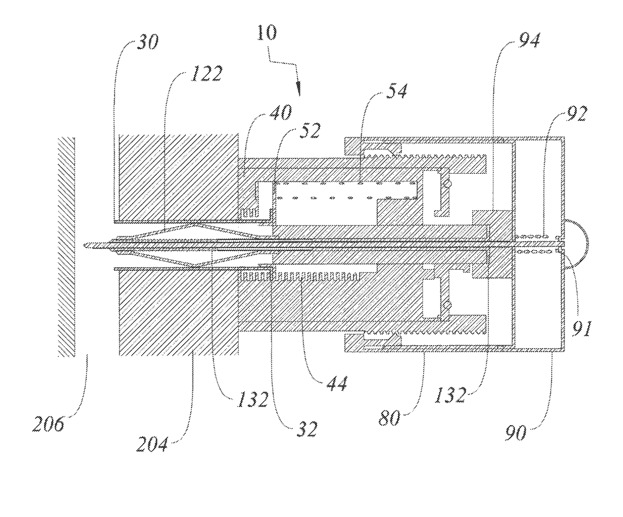
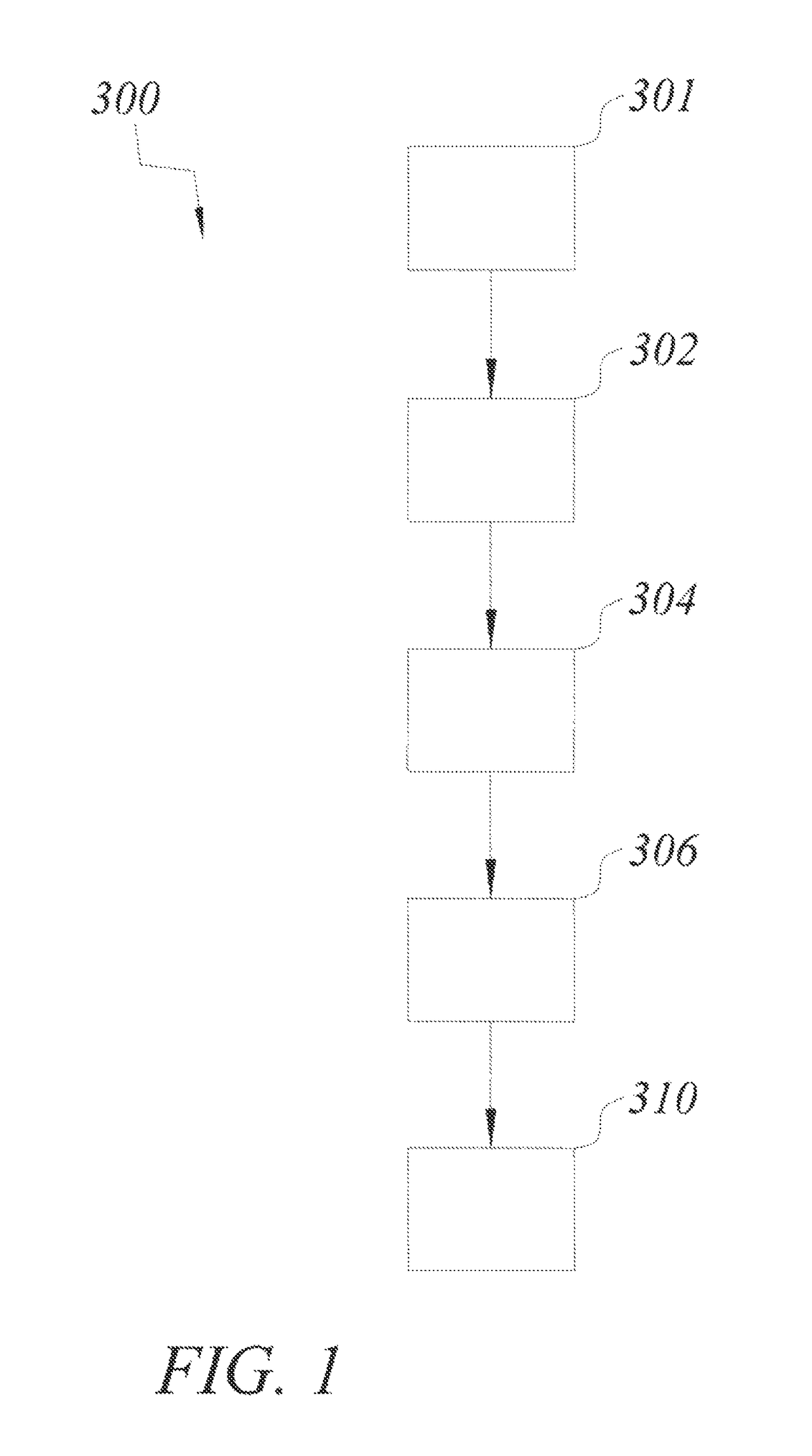
[0056]Referring to the drawings, FIG. 1 generally shows a method and workflow 300 according to an embodiment of the invention for forming and / or maintaining a percutaneous access pathway. This method and workflow 300 should be assumed to fit within standard emergency, pulmonary, and surgery protocols well known in the art (and not described here). Additionally, methods of standard needle use and safety, patient cleaning and sterilization, equipment disposal, suction setup and other standard medical practice well known in the art are not described here. The method 300 starts with providing a percutaneous access pathway at step 301. Under numerous embodiments, said access pathway includes a port and a catheter (i.e. an elongated tubular member). Under many but not all embodiments, said percutaneous access pathway also comes with an insertion device that includes a dilatational member. Under several embodiments, the dilatational member is initially positioned at least partially within ...


PUM


Abstract

An improved method and device are provided for forming and / or maintaining a percutaneous access pathway. The device generally comprises at least one of three type of components: access pathway, insertion device, and attachment device. In one embodiment, the device is used to form and / or maintain a percutaneous access pathway into the pleural cavity (i.e. tube thoracostomy). The provided assembly substantially reduces the possibility of iatrogenic infection while accessing and / or re-accessing a body space.

Description

RELATED APPLICATIONS[0001]The present application claims the benefit of U.S. Provisional Application No. 61 / 920,963 filed Dec. 26, 2013, which is incorporated herein in its entirety by reference. U.S. patent application Ser. No. 13 / 961,422, filed Aug. 7, 2013 and entitled Method and Device for Simultaneously Documenting and Treating Tension Pneumothorax and / or Hemothorax, having the same inventor as the present application, is also hereby incorporated by reference herein in its entirety.FIELD OF THE INVENTION[0002]The present disclosure relates generally to the field of medical devices, and more particularly, to devices and methods for forming and / or maintaining a percutaneous access pathway. In one application, the present invention relates to methods and devices for draining air and / or fluids from the body of a patient.BACKGROUND OF THE INVENTION[0003]A wide variety of diagnostic and / or therapeutic procedures involves the introduction of a device through a natural or artificially ...

Claims


Application Information

Patent Timeline
Patent Type & Authority Patents(United States)
IPC IPC(8): A61M1/04A61M25/06A61M27/00A61B17/34A61B17/00A61B90/40
CPCA61M27/00A61B17/34A61B17/3415A61B17/3496A61M25/0606A61M1/04A61M2210/101A61B90/40A61B2017/00809A61B2017/3409A61M2025/0024
Inventor DONALDSON, ROSS I.
Owner CRITICAL INNOVATIONS
Why Eureka
Try Eureka