Diagnostic spectrally encoded endoscopy apparatuses and systems and methods for use with same

a spectrally encoded endoscope and diagnostic technology, applied in the field of optical imaging, can solve the problems that physicians may not be able to obtain tissue information other than images, and it is difficult for physicians to characterize tissue types using doppler measurement, so as to reduce the cost of at least one of manufacture, reduce or minimize the number of optical components

Inactive Publication Date: 2019-08-06
CANON USA
View PDF77 Cites 8 Cited by
  • Summary
  • Abstract
  • Description
  • Claims
  • Application Information

AI Technical Summary

Benefits of technology

[0009]In accordance with one or more embodiments of the present disclosure, SEE apparatuses and systems, and methods and storage mediums may operate to characterize tissue type in addition to providing a morphological image to help an operator's diagnostic decision based on quantitative tissue information.
[0011]In accordance with at least one aspect of the present disclosure, tissue characterization or identification may be added to a SEE apparatus or system. At least one embodiment of the present disclosure measures tissue viscosity for identifying tissue type, which is obtained by speckle intensity autocorrelation, to efficiently identify the tissue and maintain or improve accuracy in identifying the tissue. One of the advantages of the measurement using speckle intensity autocorrelation compared to the aforementioned Doppler measurement is that using speckle intensity autocorrelation can detect random motions of scattering particles in tissue, which relates to viscosity of the tissue. Measuring viscosity can help physicians identify tissue type. Indeed, by way of a couple of examples, one or more embodiments of the present disclosure may include a SEE probe used with speckle detection, and may include a SEE probe used with speckle detection where tissue viscosity may be measured in addition to obtaining the SEE image. Thus, in one or more embodiments, intravascular laser speckle imaging (ILSI), or laser speckle imaging (LSI), may be employed to obtain viscosity measurements, and may be employed with a SEE probe.
[0025]In accordance with at least another aspect of the present disclosure, the SEE technique(s) discussed herein may be employed to reduce the cost of at least one of manufacture and maintenance of SEE devices, systems and storage mediums by reducing or minimizing a number of optical components in an interference optical system, such as an interferometer.

Problems solved by technology

A basic SEE system can only provide morphological images, and then physicians may not be able to obtain any tissue information other than images.
However, the main problem of using the Doppler measurement is that it requires continuous particle flow in one direction perpendicular to the optical axis.
Therefore, using the Doppler measurement is difficult for physicians to characterize tissue type.

Method used


Image

  • Diagnostic spectrally encoded endoscopy apparatuses and systems and methods for use with same
  • Diagnostic spectrally encoded endoscopy apparatuses and systems and methods for use with same
  • Diagnostic spectrally encoded endoscopy apparatuses and systems and methods for use with same

Examples


Embodiment Construction

[0051]One or more devices, optical systems, methods and storage mediums for characterizing tissue using a SEE technique and / or speckle detection are disclosed herein. In accordance with at least one aspect of the present disclosure, one or more devices, optical systems, methods and storage mediums discussed herein use a SEE technique with speckle detection or employ a serial time-encoded 2D imaging system with speckle detection.

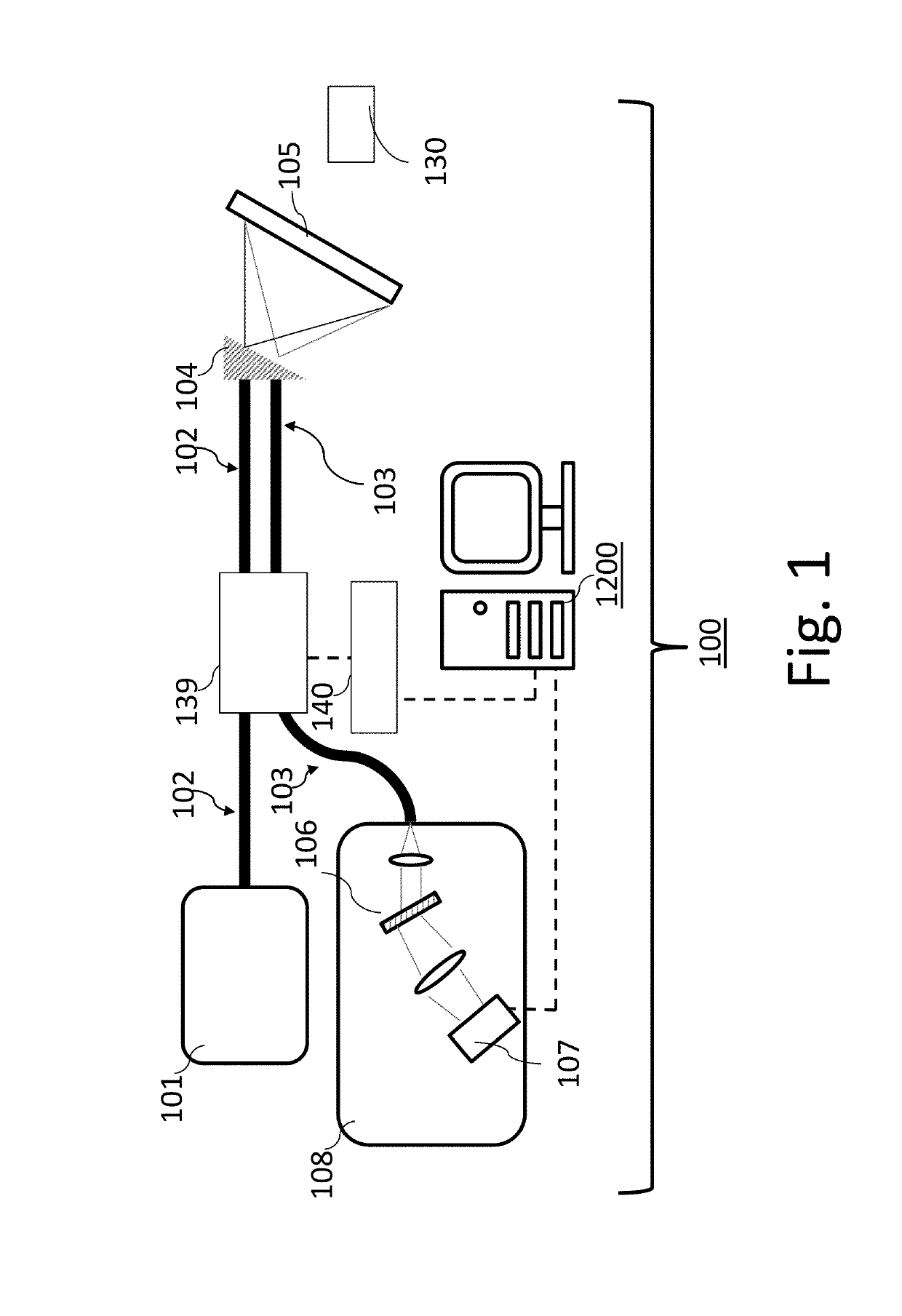
[0052]Turning now to the details of the figures, FIG. 1 shows an embodiment of a Spectrally Encoded Endoscopy (“SEE”) system 100 (also referred to herein as “system 100” or “the system 100”) which operates to utilize a SEE technique with speckle detection for optical probe applications in accordance with one or more aspects of the present disclosure. In at least one embodiment, the system 100 comprises a light source 101, an illumination fiber 102, a detection fiber 103, a motor 139, a spectrometer 108, at least one detector 107, a diffraction grating 104 tha...


PUM

PropertyMeasurementUnit
timeaaaaaaaaaa
decay timeaaaaaaaaaa
timeaaaaaaaaaa

Abstract

One or more spectrally encoded endoscopy (SEE) devices, systems, methods and storage mediums for characterizing, examining and / or diagnosing, and / or measuring viscosity of, a sample or object using speckle detection are provided. Examples of such applications include imaging, evaluating and diagnosing biological objects, such as, but not limited to, for Gastro-intestinal, cardio and / or ophthalmic applications, and being obtained via one or more optical instruments. Preferably, the SEE devices, systems methods and storage mediums include or involve speckle intensity autocorrelation function(s). One or more embodiments involve a serial time-encoded 2D imaging system with speckle detection to reconstruct images, store reconstructed images of the sample or object, and / or measure viscosity of the sample or object.

Description

CROSS-REFERENCE TO RELATED APPLICATION(S)[0001]This application claims priority to U.S. Provisional Application Ser. No. 62 / 417,084 filed 3 Nov. 2016, the content of which is incorporated herein by reference in its entirety.FIELD OF THE INVENTION[0002]The present disclosure relates generally to the field of optical imaging and more particularly to spectrally encoded endoscopy (SEE) apparatuses and systems, and methods and storage mediums for use with same. Examples of SEE applications include imaging, evaluating and characterizing / identifying biological objects or tissue, such as, but not limited to, for gastro-intestinal, cardio and / or ophthalmic applications.BACKGROUND OF THE INVENTION[0003]Spectrally encoded endoscope (SEE) is an endoscope technology which uses a broadband light source, a rotating grating and a spectroscopic detector to encode spatial information on a sample. When illuminating light to the sample, the light is spectrally dispersed along one illumination line, suc...

Claims


Application Information

Patent Timeline
Patent Type & Authority Patents(United States)
IPC IPC(8): A61B1/00A61B5/00G01N11/00A61B1/04A61B1/07
CPCG01N11/00A61B1/00009A61B1/00147A61B1/00172A61B1/07A61B5/0062A61B5/0075A61B5/0084A61B1/04G01N2011/006A61B1/000096
Inventor HOSODA, MASAKINAKAJI, HARUOWANG, ZHUO
Owner CANON USA
Why Eureka
Try Eureka