

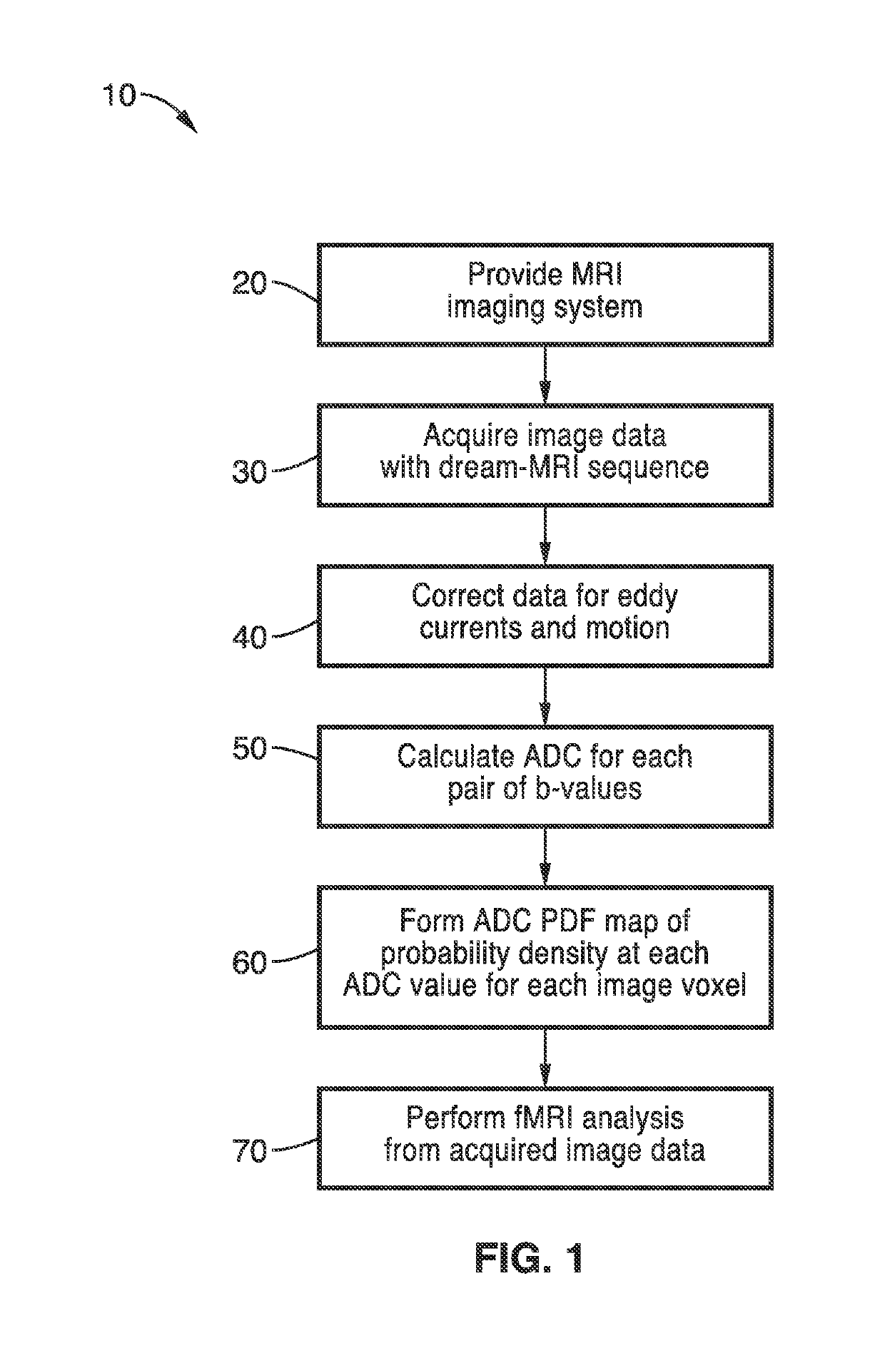
Diffusion reproducibility evaluation and measurement (DREAM)-MRI imaging methods
a reproducibility evaluation and imaging method technology, applied in the field of magnetic resonance diagnostic imaging methods, can solve the problems of not taking into account variability in adc measurement, failing to capture the complexity of tumor microstructure, etc., and achieve the effect of improving patient car
- Summary
- Abstract
- Description
- Claims
- Application Information
AI Technical Summary
Benefits of technology
Problems solved by technology
Method used
Image

Examples
example 1
[0048]Although diffusion MRI is an important cancer imaging biomarker, traditional diffusion MR techniques do not consider the variabilities in ADC measurements. ADC measurements within brain tumors vary over time and follow highly complex PDF distributions, suggesting single estimates of ADC using traditional diffusion MR approaches may not be adequate for characterizing brain tumor tissues.
[0049]The methods of the present technology overcome these issues with a new approach to diffusion MR quantification termed diffusion repeatability evaluation and measurement (DREAM)-MRI that allows for estimation of uncertainty in ADC quantification by approximating the voxel-wise ADC probability density function (PDF) distribution. The DREAM-MRI sequence is used to measure the voxel-wise ADC PDFs in water phantoms, healthy volunteers, and patients with brain tumors in Examples 2-4 respectively.
[0050]To demonstrate the Diffusion Reproducibility Evaluation and Measurement (DREAM)-MRI methods, th...
example 2
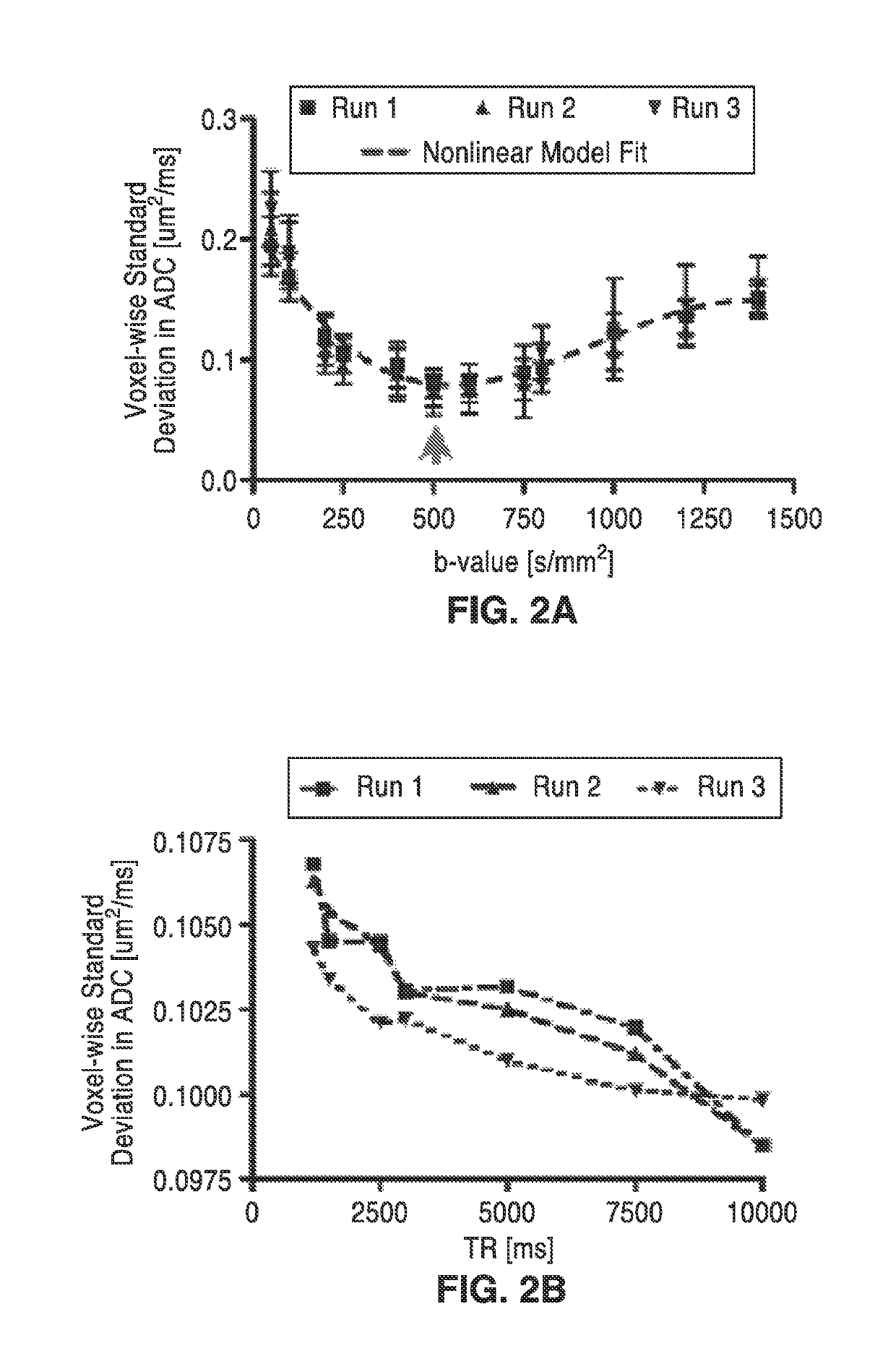
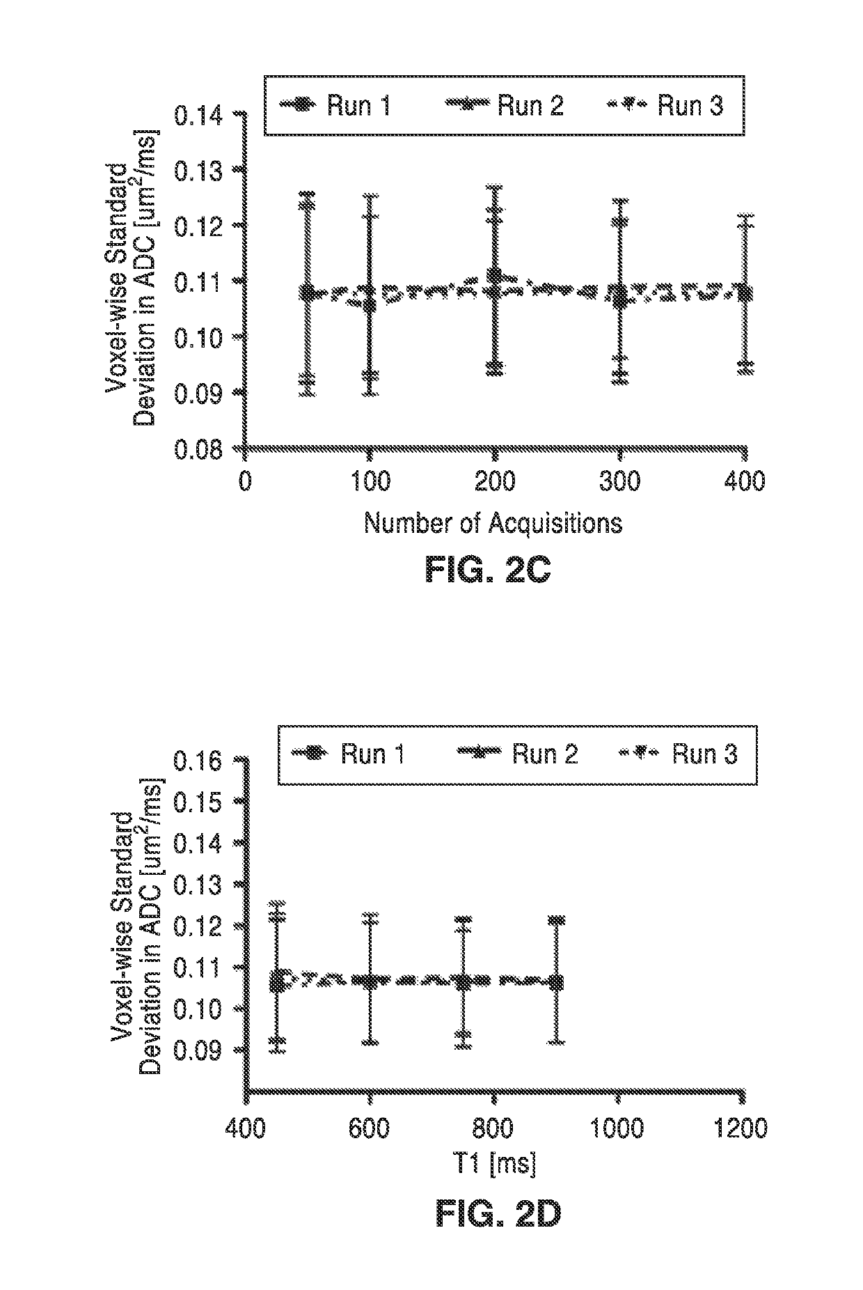
[0054]In order to further demonstrate the technology, DREAM-MRI acquisition parameters were optimized using water phantoms by minimizing ADC variability. A total of four different experiments were performed using either the American College of Radiology (ACR) water phantom (Model: J10371) or the Magphan® EMR051 Alzheimer's disease neuroimaging initiative (ADNI) phantom aimed at exploring the influence of DREAM-MR scan parameters on measurements of ADC variability. Phantom experiments for optimization of DREAM-MRI acquisition parameters are shown in FIG. 2A through FIG. 2D.
[0055]Experiment 1 consisted of an examination of the effects of b-value by averaging voxel-wise ADC variability within the isotropic area of the ACR phantom (i.e. center slices with no structures) using a single b=0 s / mm2 image and b-values from 50 to 1400 s / mm2 and TE / TR=115 ms / 1500 ms.
[0056]In order to assess the effects of b-value on ADC variability, the DREAM-MRI sequence was performed three times at a variety...
example 3
[0062]The ADC PDF characteristics for neurologically-intact healthy volunteers (n=20) were evaluated using both standard clinical MRI and DREAM-MRI acquisitions. Regions of interest for normal-appearing white matter (NAWM), normal-appearing deep gray matter (NAGMDeep), cerebrospinal fluid (CSF), and normal-appearing cortical gray matter (NAGM) were observed.
[0063]A total of 20 neurologically-intact, healthy volunteers (ages 22-35, 16 males, 4 females) were enrolled and gave their informed written consent to participate. Initially, anatomical and diffusion MRI scans were obtained in all patients using standard clinical protocols. Patients received an axial T2-weighted fast spin-echo, axial fluid-attenuated inversion recovery (FLAIR) sequence, and pre-contrast T1-weighted images. In addition to DREAM-MRI, an isotropic (3-direction) clinical DWI scan was obtained with b=0, 500, and 1000 s / mm2. Lastly, contrast-enhanced T1-weighted images (T1+C) were obtained after injection of 0.1 mmol...
PUM
 Abstract
Description
Claims
Application Information