Portable device for ex vivo stimulation of whole blood

a technology of ex vivo stimulation and portable devices, which is applied in the field of portable devices, can solve the problems of large variation in gene expression analysis from institute to institute, and the value cannot be converted to the amount of the gene in the original materials,

Active Publication Date: 2020-01-28
HITACHI CHEM CO LTD +1
View PDF12 Cites 0 Cited by
  • Summary
  • Abstract
  • Description
  • Claims
  • Application Information

AI Technical Summary

Benefits of technology

[0016]In several embodiments, there is provided a non-glass blood collection tube for the collection of blood by negative pressure comprising a non-glass tube, one or more anticoagulants, one or more leukocyte stimulating agents, a visible marker of that is activated by negative pressure, and a puncturable self-sealing cap that seals the collection tube. In several embodiments, the pressure within the collection tube is negative as compared to the pressure outside of the collection tube, thereby allowing precise volumes of blood to be collected, based on the negative pressure within the tube.

Problems solved by technology

However, due to inconsistencies in the recovery of specific subsets of leukocytes among different samples, the number of isolated leukocytes is determined for each sample and results are often expressed as the quantity of mRNA per leukocytes, not mRNA / μL blood.
Moreover, mRNA quantities may change during lengthy isolation processes.
The scientific community is facing a huge problem of institute-to-institute and experiment-to-experiment variation in gene expression analysis, because of the lack of standardization.
Although recent gene amplification technologies provide an absolute quantity of template DNA, these values cannot be converted to the amounts of the gene in the original materials, due to the lack of information of the yield of RNA recovery and the efficiency of cDNA synthesis in each sample.
Once RNA is extracted, the next step is the synthesis of cDNA, which itself can create uncertainty since existing methods do not indicate whether each RNA template creates a single copy of cDNA in each experiment.
However, the amounts of control genes are typically not consistent and may change during experiments.
Moreover, this variation presents a serious problem for clinical diagnostics, since each clinical specimen is typically analyzed at a different point in time.
It is typically very difficult to isolate pure mRNA from whole blood because whole blood contains large amounts of RNAases (from granulocytes) and non-nucleated erythrocytes.
Clin. Chem. 45:785-89), the assay procedures are labor-intensive, require several rounds of centrifugation, and involve careful handling that is essential in eliminating ribonuclease activities.

Method used


Image

  • Portable device for ex vivo stimulation of whole blood
  • Portable device for ex vivo stimulation of whole blood
  • Portable device for ex vivo stimulation of whole blood

Examples


example 1

lood Stimulation

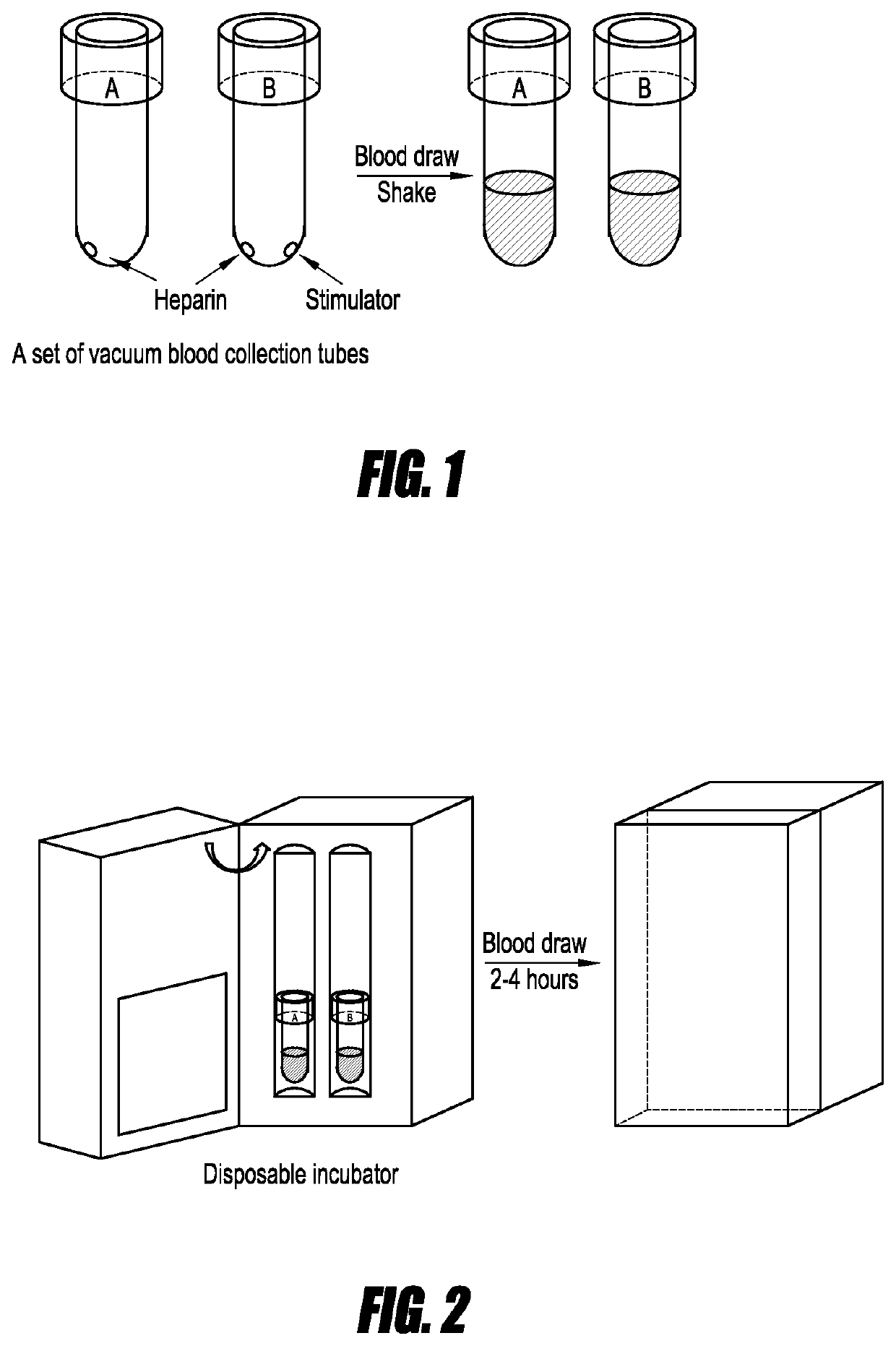
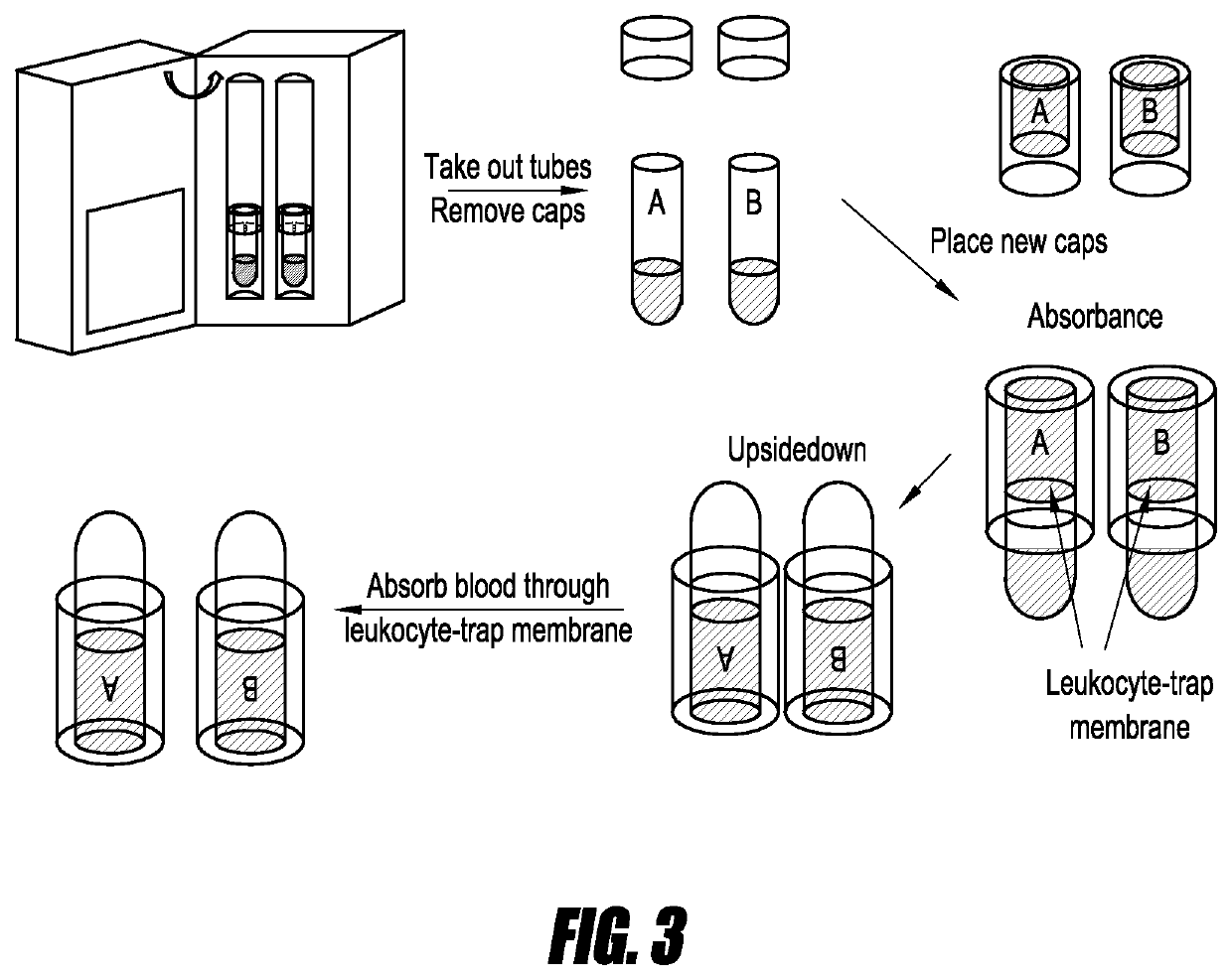
[0067]FIGS. 1-5 show methods and devices for collecting blood, processing the samples, and performing an mRNA assay on the processed blood sample in accordance with some embodiments. As illustrated in FIG. 1, tubes A and B both contain heparin prior to filling with whole blood cells. In other embodiments, heparin (or another anticoagulant) may be added to the tubes manually). Tube A is used as a control because it does not contain any stimulant. Tube B contains a dried stimulant. As above, in some embodiments, a stimulant, including an additional stimulant, is added manually. The blood is collected into the sample tubes followed by shaking or agitating the tubes to ensure that the heparin and / or stimulant mix with the collected blood.

[0068]Next, as shown in FIG. 2 a disposable incubator is used to incubate the blood samples of FIG. 1. In several embodiments, incubation is performed under physiological conditions. In some embodiments, a temperature of about 37° C. is ...

example 2

lection Tube Manufacturing and Testing

[0071]Often, blood collection tubes are made of glass, which due to manufacturing defects, exposure to fluctuations in temperature, or mishandling, can break. Given that blood samples are often collected in order to assess the health status of a patient, the blood sample may contain pathogens, chemicals, or other substances that present a danger to the individual(s) collecting and / or analyzing the blood samples. As such, in several embodiments there is provided a non-glass blood collection tube that reduces the risk of tube breakage and unintended exposure of personnel to patient blood samples. Non-glass blood collection tubes, such as those disclosed herein, are advantageous in comparison to traditional glass tubes, as collected blood samples can be stored frozen below −70° C. (e.g., for example after a stimulation protocol), but with a reduced risk of breakage during the thawing process which precedes final analysis. In several embodiments, no...

example 3

n of Manufactured Blood Collection Tubes and Multi-Well Strips

[0075]After confirming the reproducibility of manufacturing non-glass blood collection tubes, the following experiments were performed to assess whether such collection tubes perform sufficiently well as more traditional ex vivo blood stimulation methods (e.g., placement of small volumes of blood in wells and individually adding stimulating agents.

[0076]Blood collection tubes were manufactured as described above. Three tubes were stored at 4° C. (tubes PHA 4C #1, #2, and #3 in FIG. 9) or room temperature (PHA RT in FIG. 9) for 20 days. Blood was then drawn into 6 tubes: a control heparin tube, a plain heparin tube, and 4 PHA-containing tubes. The blood from the control heparin tube was stimulated (by PBS or PHA) in micro-plate tube strips (3 wells each, 60 μL blood / well). The remaining 5 tubes were incubated at 37° C. for 4 hours. Various mRNA were quantified by using SYBR green real time PCR as described previously (Mits...


PUM


Abstract

Disclosed are methods, device kits, and systems for improved quantification of mRNA from whole blood. More particularly, the devices and kites related thereto are useful for the controlled and repeatable ex vivo stimulation of whole blood.

Description

RELATED APPLICATIONS[0001]The contents of each of the priority applications listed in the accompanying Application Data Sheet is incorporated in its entirety by reference herein.FIELD OF THE INVENTION[0002]Embodiments of the present invention relate generally to portable devices capable of generating and maintaining controlled environmental conditions within the device. Several embodiments relate generally to the use of the devices for the ex vivo treatment and stimulation of whole blood.DESCRIPTION OF THE RELATED ART[0003]Research in the field of molecular biology has revealed that the genetic origin and functional activity of a cell can be deduced from the study of its ribonucleic acid (RNA). This information may be of use in clinical practice, to diagnose infections, to detect the presence of cells expressing oncogenes, to detect familial disorders, to monitor the state of host defense mechanisms and to determine the HLA type or other marker of identity. RNA exists in three funct...

Claims


Application Information

Patent Timeline
Patent Type & Authority Patents(United States)
IPC IPC(8): A01N1/02A61B5/15A61K35/14
CPCA61B5/150206A01N1/0284A01N1/0252A61B5/1411A61B5/15003A61B5/150305A61B5/150389A61B5/150755A61B5/153A61B5/154A61K35/14
Inventor MITSUHASHI, MASATOMURAKAMI, TAKU
Owner HITACHI CHEM CO LTD
Why Eureka
Try Eureka