
Method and apparatus for treating valvular disease

a valvular disease and treatment method technology, applied in the field of valvular stenosis, can solve the problems of significant risk of death or serious complications, significant surgical risks, and wide application rang

Active Publication Date: 2020-08-11
CARDIAWAVE
View PDF42 Cites 0 Cited by
  • Summary
  • Abstract
  • Description
  • Claims
  • Application Information

AI Technical Summary

Problems solved by technology

It is thus a major operation that conveys significant risk of death or serious complications.
Moreover, a large class of older patients, as well as those who are frail and / or have multiple comorbidities, face significantly higher surgical risks and are thus excluded from the scope of application of this method.
However, these catheter-based approaches are only applicable to selected groups of patients and still involve significant risk of death or serious complications.
Even when a patient fulfils the conditions to receive a replacement valve by open or percutaneous surgery, both mechanical and tissue-based replacement heart valves present significant drawbacks.
Mechanical valves are made from pyrolytic carbon and require a life-time treatment of warfarin anticoagulant, with an accompanying risk of bleeding.
While such bleeding events are rare, they are often fatal.
Over this time the valve will likely be degenerating to the point of requiring replacement, which again carries a significant risk of death.
Moreover, tissue valves are also subject to valvular diseases; in particular, they may also develop fibro-calcific degeneration requiring an early replacement.

Method used


Image

  • Method and apparatus for treating valvular disease
  • Method and apparatus for treating valvular disease
  • Method and apparatus for treating valvular disease

Examples


Embodiment Construction

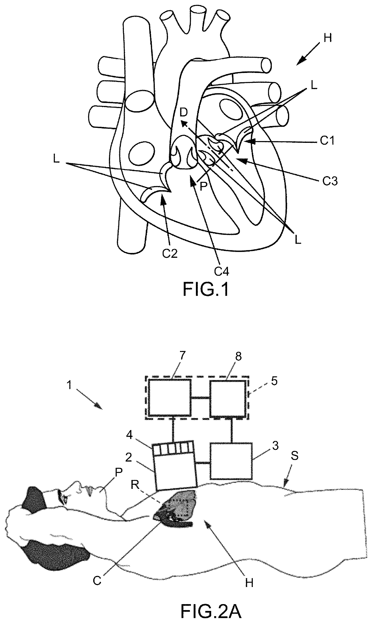
[0070]FIG. 1 illustrates a heart H of a patient which is a mammalian, for instance a human. The heart comprises four cardiac valves C1, C2, C3, C4 that determine the pathway of blood flow through the heart: the mitral valve C1, the tricuspid valve C2, the aortic valve C3 and the pulmonary valve C4.

[0071]Each cardiac valve C allows blood to flow in only one direction through the heart H by opening or closing incumbent on differential blood pressure on each side of the valve.

[0072]More precisely, each cardiac valve C comprises leaflets L, also called cusps, which are thin tissue layers that are able to be closed together, to seal the valve and prevent backflow, and pushed (i.e. bended) open to allow blood flow. The mitral valve C1 usually has two leaflets L, whereas the three others cardiac valves C2, C3, C4 usually have three leaflets L (only two leaflets are show on FIG. 1 for each cardiac valve). The leaflets are fixed to an annulus of the cardiac valve C. The annulus is a ring com...


PUM


Abstract

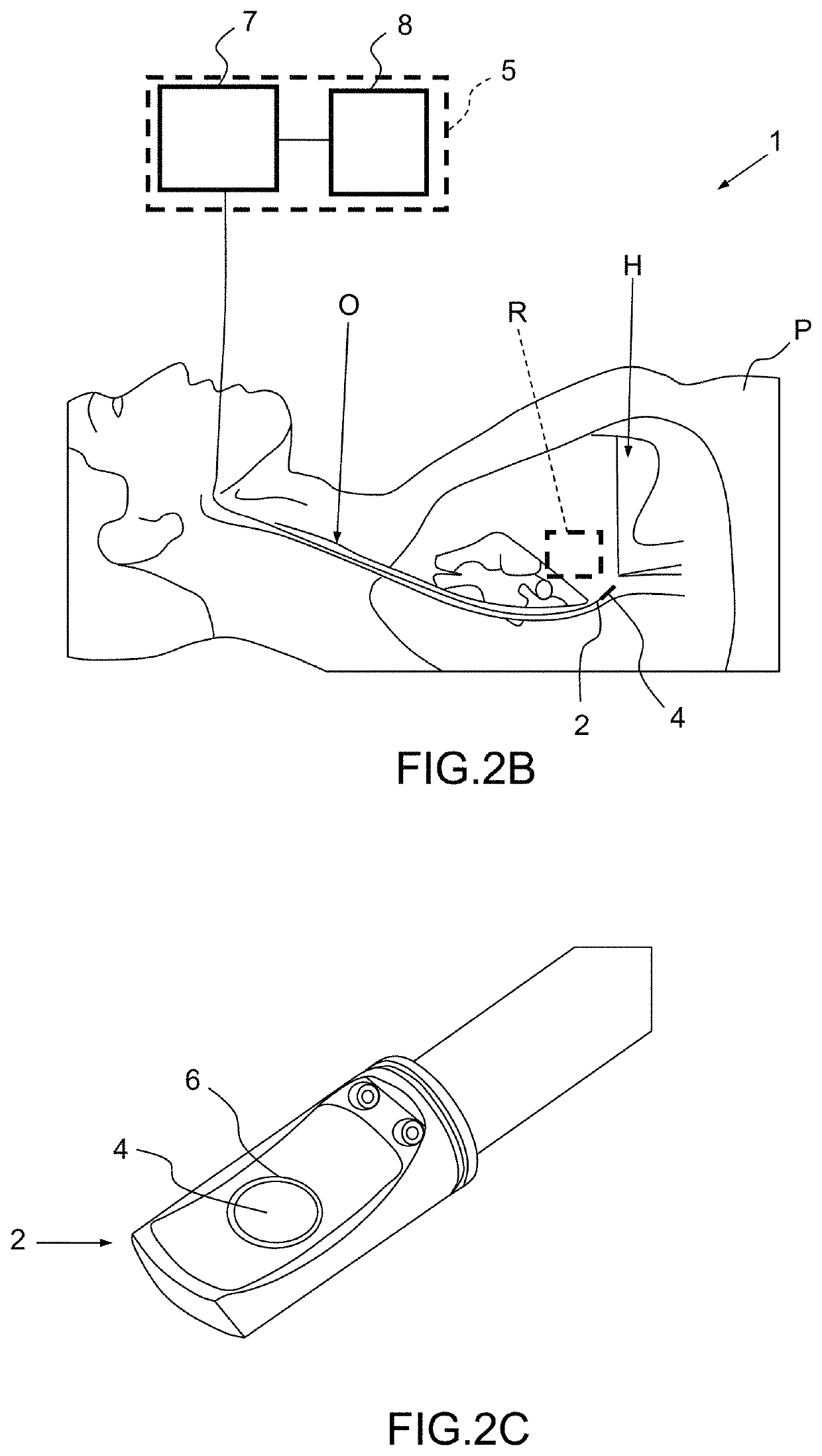
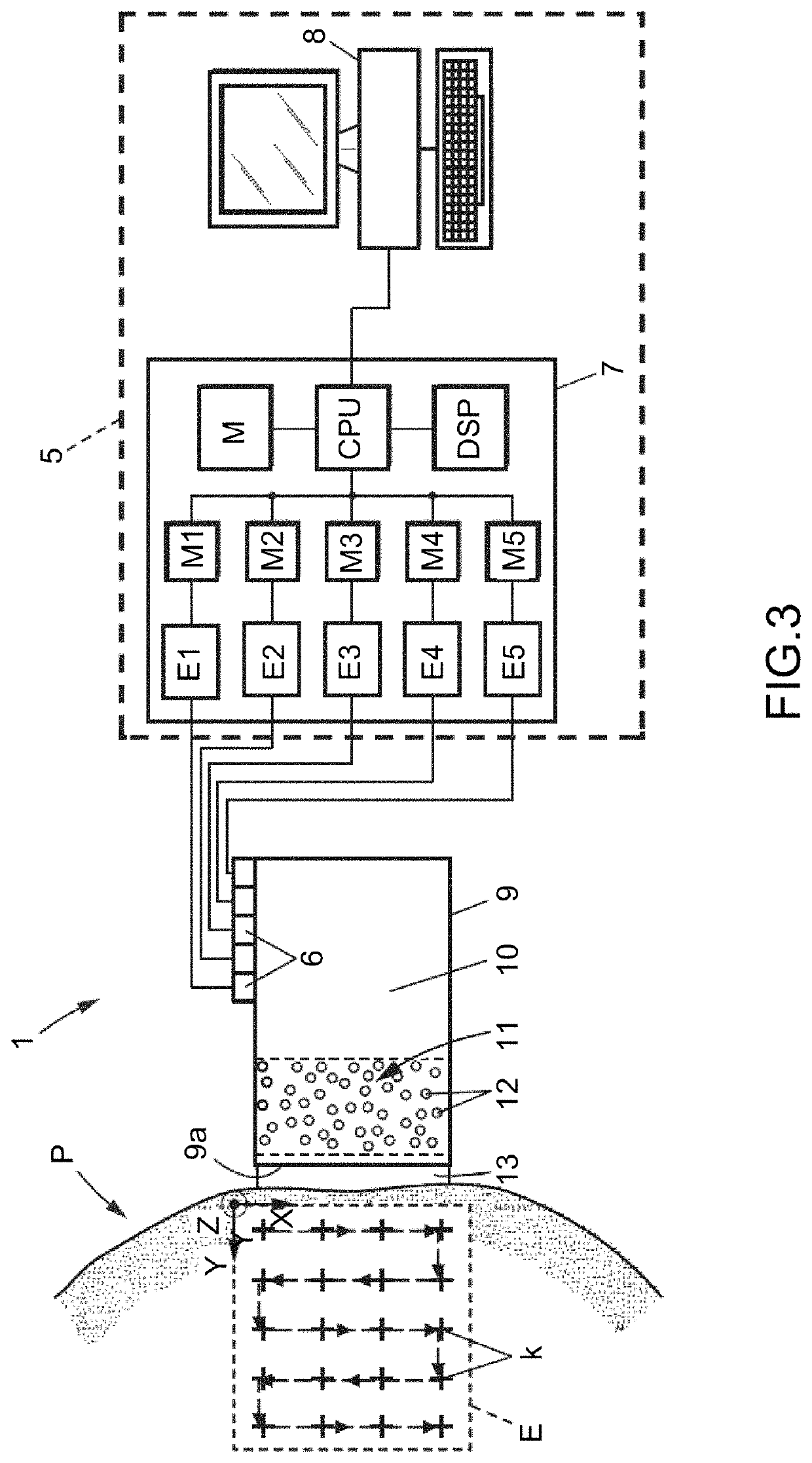
An apparatus for treating or preventing a valvular disease comprises: a ultrasound probe located externally to a heart of patient, able to produce ultrasound waves focused inside the heart and suitable to generate, at a focal spot, a pressure sufficient to result in cavitation, an imaging device for mapping in real time a treatment region of a cardiac valve of the patient, the treatment region comprising at least one leaflet of the cardiac valve, a controller configured for driving the ultrasound probe to emit a sequence of focused ultrasound waves, the controller being further configured for steering the focused ultrasound waves so as to scan the entire treatment region to soften the tissues of the treatment region. A method for treating or preventing valvular disease, carried out using the apparatus is also provided.

Description

CROSS-REFERENCE TO RELATED APPLICATIONS[0001]This application is a National Stage of International patent application PCT / IB2016 / 000523, filed on Apr. 1, 2016, which claims priority to U.S. provisional application No. 62 / 142,096, filed on Apr. 2, 2015, the disclosures of which are incorporated by reference in their entirety.FIELD OF THE INVENTION[0002]The instant invention relates to methods and apparatuses for treatment or prevention of valvular diseases, in particular valvular stenosis.BACKGROUND OF THE INVENTION[0003]Valvular stenosis, also called heart valve disease or narrowed valve, occurs when tissues forming a cardiac valve leaflets become stiffer thereby narrowing the valve opening and reducing the amount of blood that can flow through it.[0004]Valvular stenosis may occur in any of the four valves of the heart: the aortic valve, the mitral valve, the tricuspid valve or the pulmonic valve.[0005]One of the known causes of valvular stenosis is a fibro-calcific degeneration of ...

Claims


Application Information

Patent Timeline
Patent Type & Authority Patents(United States)
IPC IPC(8): A61B8/06A61B8/12A61B8/04A61N7/00A61F2/24A61B34/30A61B90/00A61B5/00A61B8/00A61B17/00A61B5/0215A61N7/02A61B8/08A61B34/20
CPCA61B8/12A61B8/06A61B8/0883A61N7/02A61B8/04A61B2090/376A61B2034/2063A61B8/4483A61B2017/00398A61B34/30A61B2034/2065A61N2007/0095A61N2007/0039A61B8/445A61B2090/3784A61N2007/0052A61B8/488A61B2576/023A61N2007/0086A61B5/0215A61N2007/0004A61F2/24A61B2090/378A61B2090/3782A61B8/485A61N7/00A61B2090/374A61B5/0053G16H30/40A61B2017/22098A61B17/225A61B17/22004
Inventor MESSAS, EMMANUELPERNOT, MATHIEUTANTER, MICKAELVILLEMAIN, OLIVIER
Owner CARDIAWAVE
Learn More