
In vitro epithelial models comprising lamina propria-derived cells

a technology of epithelial cells and lamina propria, which is applied in the field of in vitro epithelial models comprising lamina propria-derived cells, can solve the problems of large limitations of models, limited cellular heterogeneity, and complex 3-dimensional (3d) architecture of explant cultures, and achieve the effect of reducing inflammation in epithelial regions

Active Publication Date: 2020-11-10
EMULATE INC
View PDF17 Cites 2 Cited by
  • Summary
  • Abstract
  • Description
  • Claims
  • Application Information

AI Technical Summary

Problems solved by technology

However, these models have significant limitations.
Explant cultures, which have organotypic properties such as complex 3-dimensional (3D) architecture and cellular heterogeneity are limited in part by their lack of reproducibility of growing conditions between laboratories and their short-term nature.
For example, a selectively-permeable membrane can allow passage of a fluid (e.g., liquid and / or gas), nutrients, wastes, cytokines, and / or chemokines from one side of the membrane to another side of the membrane, but does not allow whole living cells to pass therethrough.

Method used


Image

  • In vitro epithelial models comprising lamina propria-derived cells
  • In vitro epithelial models comprising lamina propria-derived cells
  • In vitro epithelial models comprising lamina propria-derived cells

Examples


example 1

Lung On-Chip: Evaluation of Epithelial Cells in the Upper Compartment

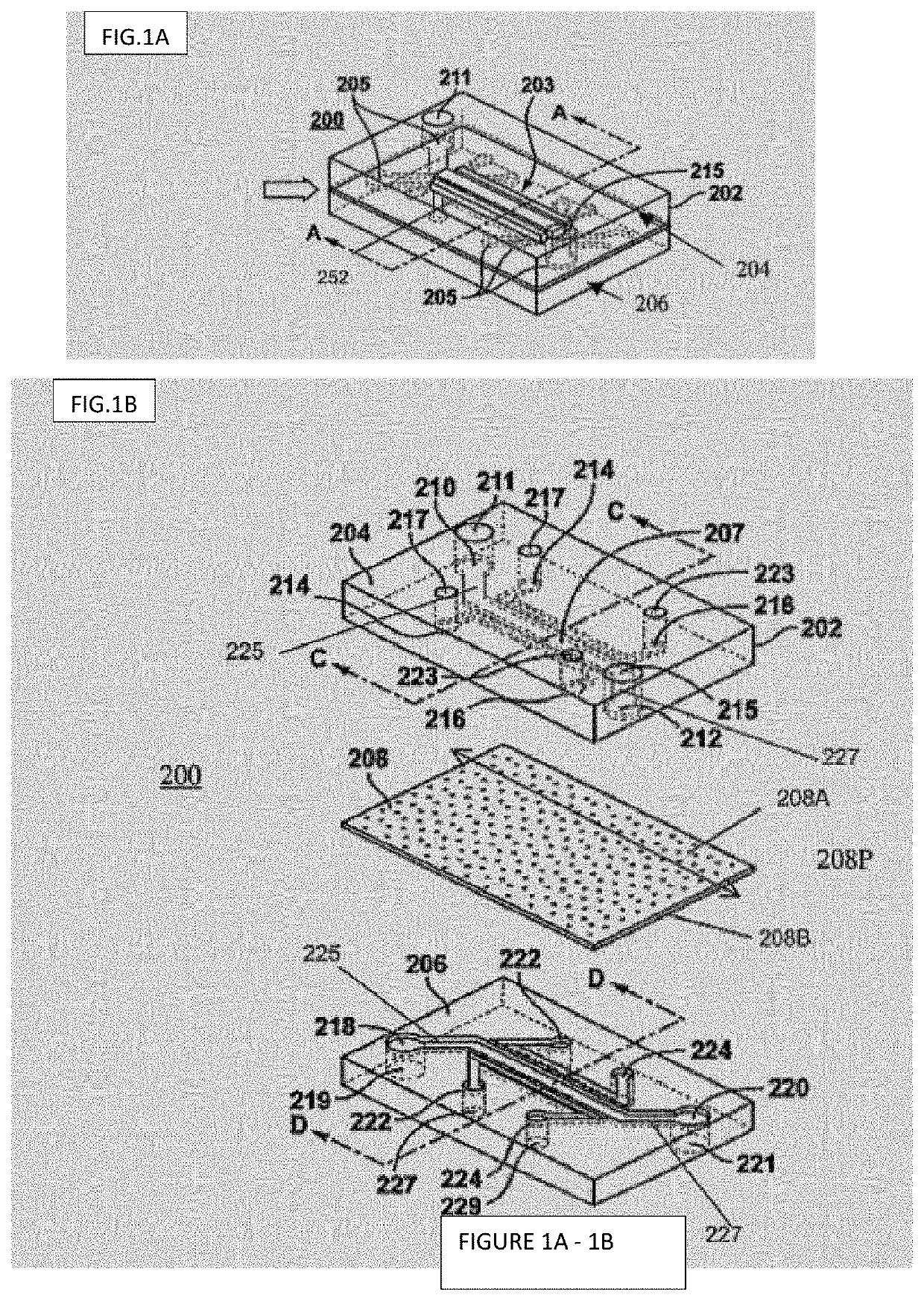
[0233]In one embodiment, an exemplary timeline was used for preparing, seeding, and evaluating an Alveolus Lung-On-Chip. FIG. 5 shows an exemplary schematic of one embodiment of an experimental timeline where Gel preparation is on Day-2; ECM is added to microchannels on Day-1; Cell seeding is on Day 0; Air-Liquid Interface is established by Day 5; Stretch of 4% begins on Day 9; Stretch of 10% begins on Day 12 which may continue up to Day 15. Cell seeding included fibroblast cells within a gel, alveolar epithelial cells overlaying the gel in the upper channel, and endothelial cells in the lower channel. Incorporation of more physiologically relevant ECM (Elastin).

[0234]In some embodiments, mechanical stretch was providing using vacuum applied to vacuum channels for moving the membrane within the chip simulating in vivo breathing movements. In some embodiments, read-outs, i.e. evaluation, includes but is not limited ...

example 2

Lung On-Chip: Evaluation of Stromal Cells in the Stromal Compartment

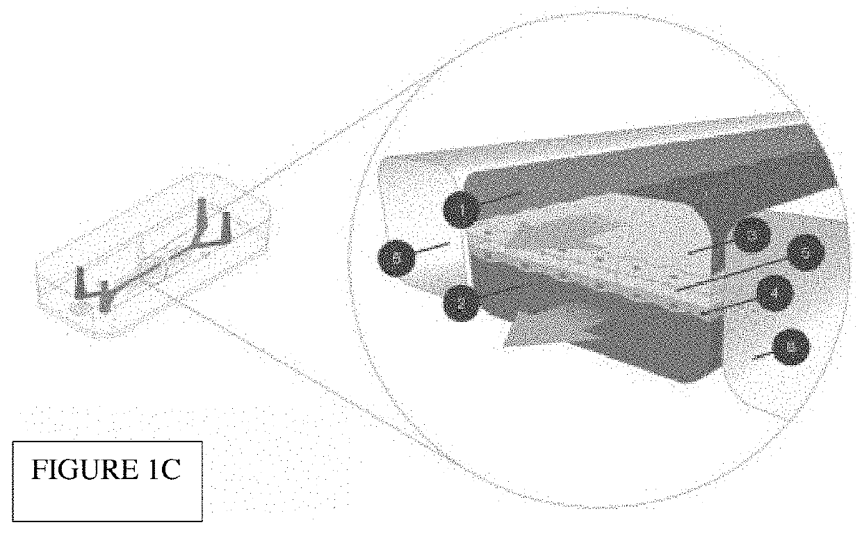
[0245]Human primary lung fibroblasts have been incorporated within the 3D stromal compartment of the chip, see highlighted area on-chip shown in FIG. 4C. Incorporation of Lung Fibroblasts On-Chip rhodamine phalloidin (PHDR1)Alexa Fluor® 488 Phalloidin binds to F-actin proteins.

[0246]FIGS. 13A-B Show exemplary micrographs demonstrating the presence of lung fibroblasts. FIG. 13A lung fibroblasts stained for Phalloidin (pink) and Nuclei (blue). FIG. 13B Phalloidin (pink) and Type I-Like cells (green). White Bar=100 um.

[0247]FIGS. 14A-C Show exemplary micrographs demonstrating assessment of Fibroblast Viability growing on-chip. FIG. 14A Maximum intensity projection of Z-Stack. FIG. 14B Live / Dead. FIG. 14C Phase Contrast (left) Phase Contrast Merge with Live / Dead. Live / dead staining shows high percentage of cell surviving on chip on day 15 post-seeding. Dead fibroblasts: rounded morphology and red nuclei. Live fibroblast...

example 3

Lung On-Chip: Evaluation of the Lower Vascular Compartment

[0251]FIG. 17 Shows exemplary micrographs demonstrating microvascular endothelial cells expressing typical endothelial cells markers and covering the entire surface of vascular channel at 2 post seeding. Left to right: PECAM-1; VE-Cadherin; and VWF.


PUM

PropertyMeasurementUnit
thicknessaaaaaaaaaa
thicknessaaaaaaaaaa
thicknessaaaaaaaaaa

Abstract

An in vitro microfluidic “organ-on-chip” is described herein that mimics the structure and at least one function of specific areas of the epithelial system in vivo. In particular, a multicellular, layered, microfluidic culture is described, allowing for interactions between lamina propria-derived cells and the associated tissue specific epithelial cells and endothelial cells. This in vitro microfluidic system can be used for modeling inflammatory tissue, e.g., autoimmune disorders involving epithelia and diseases involving epithelial layers. These multicellular, layered microfluidic “organ-on-chip”, e.g. “epithelia-on-chip” further allow for comparisons between types of epithelia tissues, e.g., lung (Lung-On-Chip), bronchial (Airway-On-Chip), skin (Skin-On-Chip), cervix (Cervix-On-Chip), blood brain barrier (BBB-On-Chip), etc., in additional to neurovascular tissue, (Brain-On-Chip), and between different disease states of tissue, i.e. healthy, pre-disease and diseased areas. Additionally, these microfluidic “organ-on-chips” allow identification of cells and cellular derived factors driving disease states in addition to drug testing for reducing inflammation effecting epithelial regions.

Description

FIELD OF THE INVENTION[0001]An in vitro microfluidic “organ-on-chip” is described herein that mimics the structure and at least one function of specific areas of the epithelial system in vivo. In particular, as multicellular, layered, microfluidic culture is described, allowing for interactions between lamina propria-derived cells and epithelial cells and endothelial cells. This in vitro microfluidic system can be used liar modeling inflammatory tissue, e.g. autoimmune disorders involving epithelia and diseases involving epithelial layers. These multicellular, layered microfluidic “organ-on-chip”, e.g. “epithelia-on-chip” further allow for comparisons between types of epithelia tissues, e.g., lung (Lung-On-Chip), bronchial (Airway-On-Chip), skin (Skin-On-Chip), cervix (Cervix-On-Chip), blood brain barrier (BBB-On-Chip), etc., in additional to neurovascular tissue, (Brain-On-Chip), and between different disease states of tissue, i.e. healthy, pre-disease and diseased areas. Additiona...

Claims


Application Information

Patent Timeline
Patent Type & Authority Patents(United States)
IPC IPC(8): B01L3/00C12M1/00C12M3/06C12M1/42G01N35/00G01N33/48G01N33/50C12N5/00
CPCB01L3/502753C12M35/08C12M29/04B01L3/502715G01N33/5044C12M23/16C12N5/0075C12N5/0018
Inventor KERNS, S. JORDANBARRILE, RICCARDOHAMILTON, GERALDINEKARALIS, CATHERINELEVNER, DANIELLUCCHESI, CAROLINAVARONE, ANTONIOVILLENAVE, REMI
Owner EMULATE INC
Learn More