Detection and analysis method for urine-modified nucleoside based on surface-enhanced resonance Raman spectroscopy
a raman spectroscopy and tumor marker technology, applied in the field of biomedical optics, can solve the problems of difficult to achieve ideal detection effect, complicated and interfered components in urine, and weak raman spectroscopy signal of conventional raman spectroscopy, so as to eliminate fluorescent background interference and eliminate experimental system errors
- Summary
- Abstract
- Description
- Claims
- Application Information
AI Technical Summary
Benefits of technology
Problems solved by technology
Method used
Image
Examples
application example 1
Clinical Application Example 1
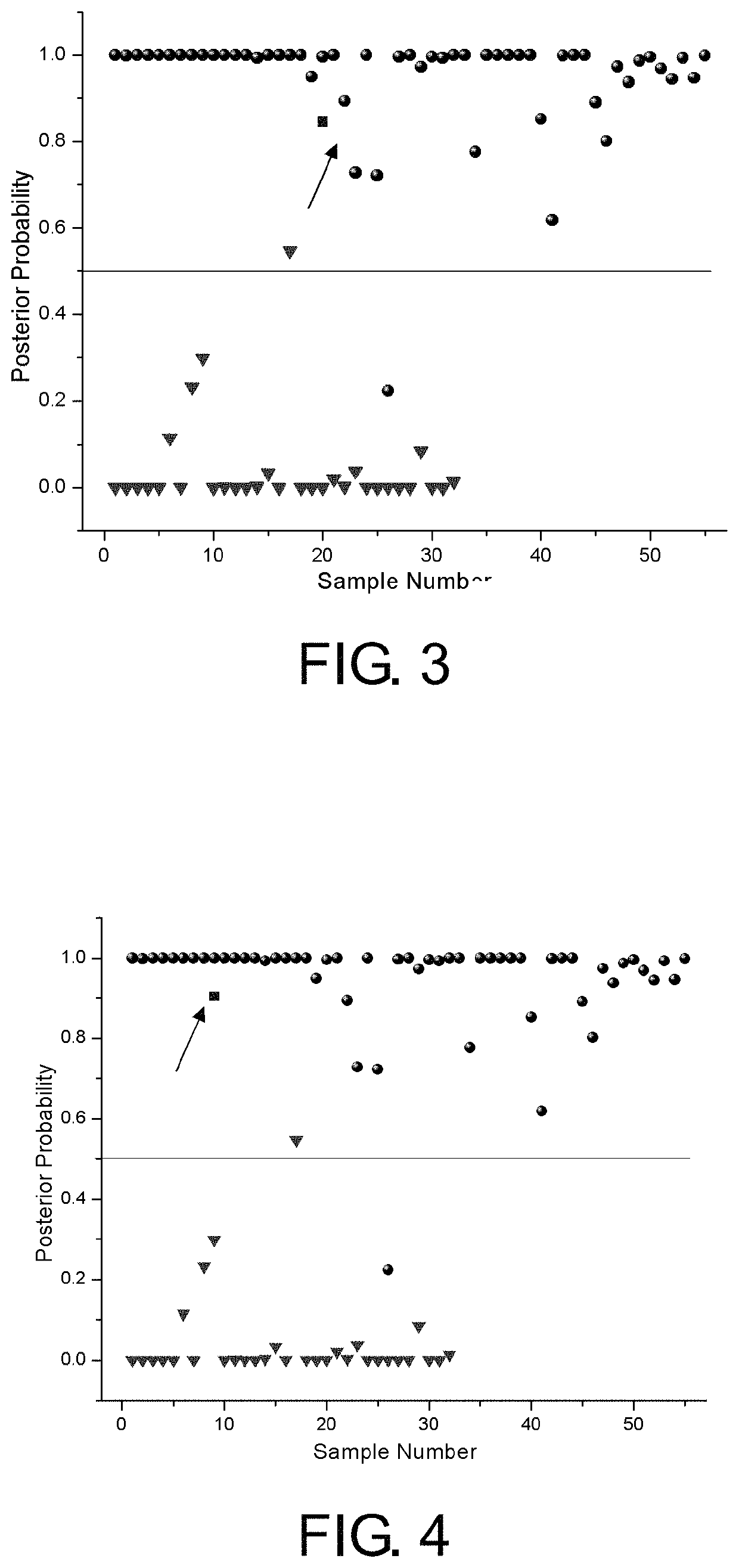
[0059]Liao X X, male, 52 years old, pathological diagnosis of myeloid highly and moderately differentiated squamous cell carcinoma. Cancer tissue covers the whole layer. Cancer embolus is seen in the blood vessel, staged as T4, N1 and Mx. Double-blind detection is conducted by the present invention. The posterior probability P=0.845 for the SERRS of the urine-modified nucleoside of the patient is calculated through the PLS-DA model. The calculation result shows that the patient is judged as a patient with esophagus cancer. FIG. 3 is the diagnosis result of the application example 1. The figure shows the posterior probability corresponding to the surface-enhanced Raman scattering of the urine-modified nucleoside of each sample. In the figure, circular points represent the patients with esophagus cancer, triangular pointed points represent normal healthy persons, and a square point represents a to-be-identified patient.
[0060]In FIG. 3, the SERRS of the ur...
application example 2
Clinical Application Example 2
[0062]Zou X X, male, 61 years old, pathological diagnosis of fungating type highly and moderately differentiated squamous cell carcinoma. Cancer tissue covers the muscular layer. Cancer embolus is seen in the vessel, staged as T2, N0 and Mx. Double-blind detection is conducted by the present invention. The posterior probability P=0.904 for the SERRS of the urine-modified nucleoside of the patient is calculated through the PLS-DA model. The calculation result shows that the patient has cancer. FIG. 4 is the diagnosis result of the application example 2. The figure shows the posterior probability corresponding to the surface-enhanced Raman scattering of the urine-modified nucleoside of each sample. In the figure, circular points represent patients with esophagus cancer, triangular pointed points represent the normal healthy persons, and a square point represents a to-be-identified patient.
PUM
| Property | Measurement | Unit |
|---|---|---|
| molar concentration | aaaaa | aaaaa |
| time | aaaaa | aaaaa |
| time | aaaaa | aaaaa |
Abstract
Description
Claims
Application Information
 - R&D
- Intellectual Property
- Life Sciences
- Materials
- Tech Scout
- Unparalleled Data Quality
- Higher Quality Content
- 60% Fewer Hallucinations
Browse by: Latest US Patents, China's latest patents, Technical Efficacy Thesaurus, Application Domain, Technology Topic, Popular Technical Reports.
© 2025 PatSnap. All rights reserved.Legal|Privacy policy|Modern Slavery Act Transparency Statement|Sitemap|About US| Contact US: help@patsnap.com


