Dixon-type water/fat separation MR imaging

a technology of water/fat separation and dixon tse, applied in the field of magnetic resonance imaging, can solve the problems of reducing the sampling efficiency of the signal, so as to achieve high signal sampling efficiency and low bandwidth. the effect of high sampling efficiency

Active Publication Date: 2021-06-22
KONINKLJIJKE PHILIPS NV
View PDF18 Cites 0 Cited by
  • Summary
  • Abstract
  • Description
  • Claims
  • Application Information

AI Technical Summary

Benefits of technology

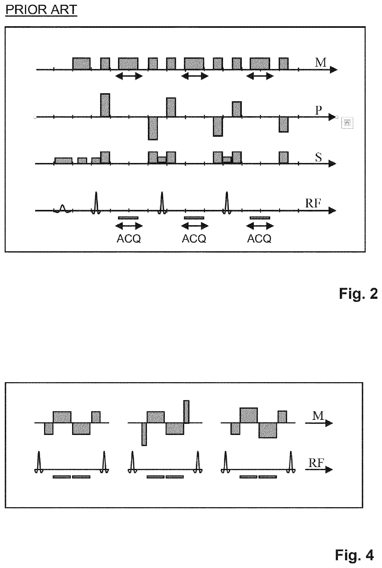
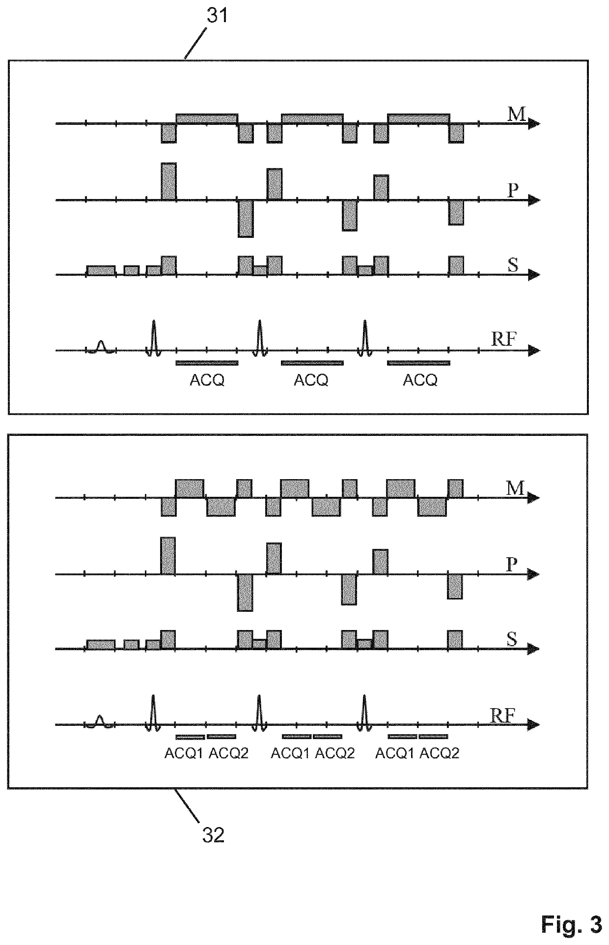
[0020]More specifically, the echo signals generated by the first imaging sequence are acquired using the unipolar readout magnetic field gradients having a first gradient strength, and the pairs of echo signals generated by the second imaging sequence are acquired using the bipolar readout magnetic field gradient having a second gradient strength which is larger than the first gradient strength. Simultaneously, the first echo signals are acquired using a signal receiving bandwidth which is smaller than the signal receiving bandwidth used for the acquisition of the second echo signals. The whole acquisition is thus split up into two, usually interleaved sub-acquisitions, performed with a low-bandwidth sub-sequence (the first imaging sequence) and a high-bandwidth sub-sequence (the second imaging sequence), respectively. In the low-bandwidth sub-sequence, a high signal sampling efficiency is achieved by sampling the first echo signals during most of the interspacing between the refocusing RF pulses. This low-bandwidth and high sampling efficiency yields a high SNR. The first imaging sequence with its single-echo readout is actually the same as a standard TSE sequence and thus provides the same sampling efficiency. The second imaging sequence achieves only a lower sampling efficiency and provides a lower SNR per echo, since it has to cover the same gradient integral in about half the time. However, by sampling a pair of echo signals, it achieves a similar, high sampling efficiency and SNR as the first imaging sequence.
[0021]The MR image reconstructed from the echo signals acquired according to the invention benefits from the higher sampling efficiency and higher SNR of both imaging sequences. This applies to a reconstructed in-phase MR image (including both water and fat signal contributions as a conventional spin echo image) as well as to a water (or fat-suppressed) MR image.
[0028]reconstructing a MR image from the acquired echo signals, wherein each pair of echo signals is combined into a virtual echo signal, wherein signal contributions from water protons and fat protons are separated by a one-point Dixon technique using the virtual echo signals. This variant may be applied in combination with any other feature of the method of the invention disclosed herein, in particular regarding the concept of FID artifact suppression described below. The single-point method has basically the same advantages as discussed above (high SNR, good sampling efficiency). However, it relates only to single-echo instead of dual-echo Dixon imaging.
[0033]From S1 and S3*, P02 can be derived. Since it is expected to be spatially smooth, it can be filtered to reduce noise. S13 is then obtained byS13=S1+S*3P02,for example. Optionally, P2 can be derived from S2, and S13 can be multiplied with P2*.
[0037]In the second imaging sequence, only the readout magnetic field gradient differs from the first imaging sequence. Its zeroth moment is usually zero both at the echo times of the pair of echo signals and at the time of the subsequent refocusing RF pulse. Its first moment is non-zero at the echo time of the first of the pair of echo signals while it is zero at the echo time of the second of the pair of echo signals. In this way, differences between the two single-echo MR images reconstructed from the pair of echo signals can be exploited for an improved flow compensation. For example, a higher signal amplitude at a given voxel position in the second single-echo MR image than in the first single-echo MR image can be attributed to flow-related intra-voxel dephasing and can be compensated by considering the signal amplitude in the second single-echo MR image only.

Problems solved by technology

However, constraints on the echo times may actually render dual-echo acquisitions slower than triple-echo acquisitions.
A drawback of the conventional approach is that, in comparison to standard (non-Dixon) TSE sequences, a higher receive bandwidth is required for a given echo spacing in Dixon TSE sequences.
However, longer or more echo trains are required in this case.
This results in less coverage and more blurring in the reconstructed MR images, or in longer scan times. Furthermore, FID artifacts may be a problem with the conventional approach.

Method used


Image

  • Dixon-type water/fat separation MR imaging
  • Dixon-type water/fat separation MR imaging
  • Dixon-type water/fat separation MR imaging

Examples


Embodiment Construction

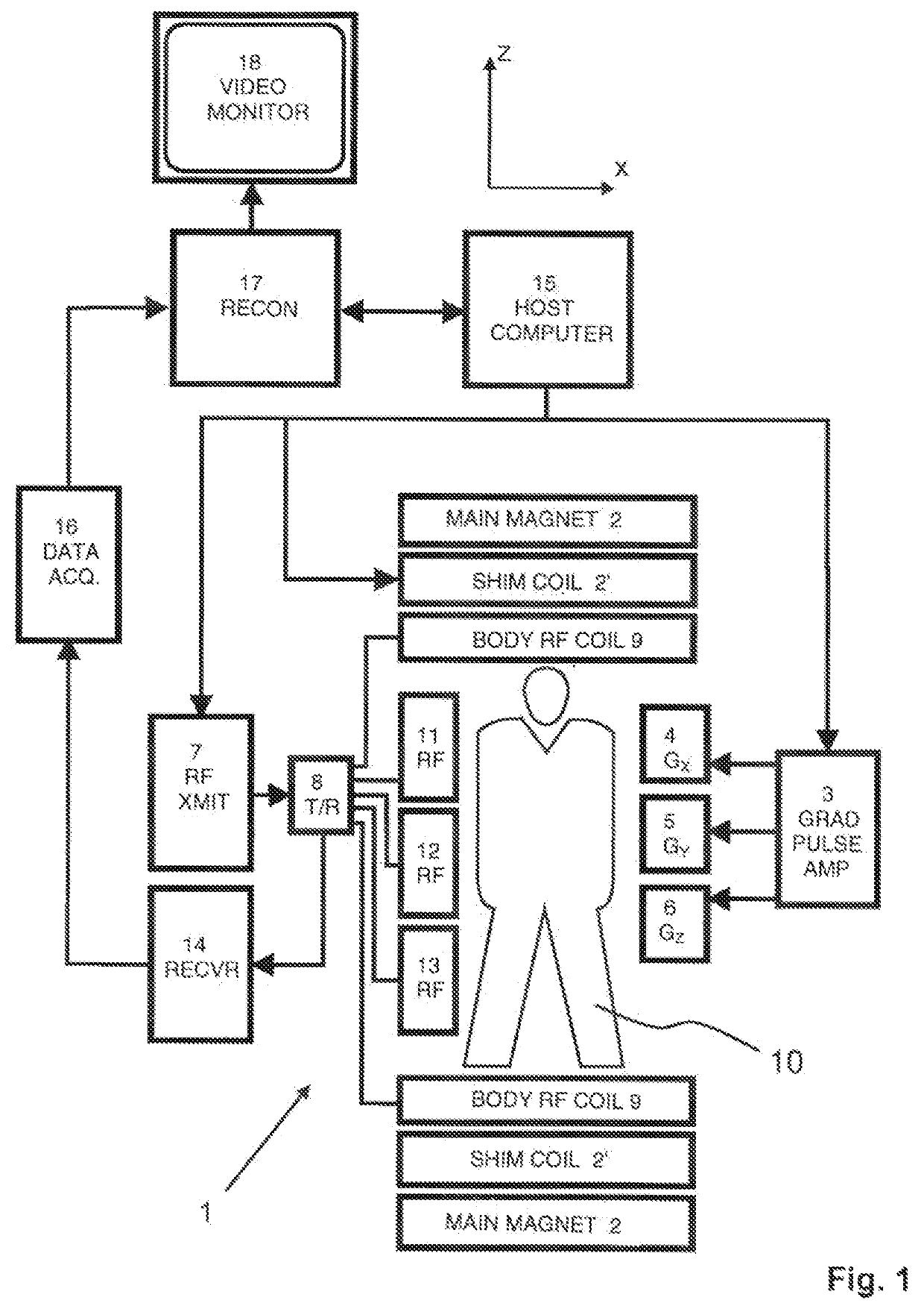
[0046]With reference to FIG. 1, a MR device 1 is shown as a block diagram. The device comprises superconducting or resistive main magnet coils 2 such that a substantially uniform, temporally constant main magnetic field B0 is created along a z-axis through an examination volume. The device further comprises a set of (1st, 2nd, and—where applicable—3rd order) shimming coils 2′, wherein the current flow through the individual shimming coils of the set 2′ is controllable for the purpose of minimizing B0 deviations within the examination volume.

[0047]A magnetic resonance generation and manipulation system applies a series of RF pulses and switched magnetic field gradients to invert or excite nuclear magnetic spins, induce magnetic resonance, refocus magnetic resonance, manipulate magnetic resonance, spatially and otherwise encode the magnetic resonance, saturate spins, and the like to perform MR imaging.

[0048]More specifically, a gradient pulse amplifier 3 applies current pulses to sele...


PUM


Abstract

A method of Dixon-type MR imaging includes subjecting the object (10) to a first imaging sequence (31) including a series of refocusing RF pulses. A single echo signal is generated in the time interval between two consecutive refocusing RF pulses. The first echo signals from the object (10) are acquired at a first receive bandwidth using unipolar readout magnetic field gradients. The object (10) is further subject to a second imaging sequence (32), which includes a series of refocusing RF pulses. A pair of second echo signals is generated in each time interval between two consecutive refocusing RF pulses. The pairs of second echo signals from the object (10) are acquired at a second receive bandwidth using bipolar readout magnetic field gradients. The second receive bandwidth is higher than the first receive bandwidth. Signal contributions from water protons and fat protons are separated and an MR image is reconstructed.

Description

CROSS REFERENCE TO RELATED APPLICATIONS[0001]This application is a U.S. national phase application of International Application No. PCT / EP2017 / 063326, filed on Jun. 1, 2017, which claims the benefit of EP Application Serial No. 16172658.3 filed on Jun. 2, 2016 and is incorporated herein by reference.FIELD OF THE INVENTION[0002]The invention relates to the field of magnetic resonance (MR) imaging. It concerns a method of MR imaging of a portion of a body placed in the examination volume of a MR device. The invention also relates to a MR device and to a computer program to be run on a MR device.BACKGROUND OF THE INVENTION[0003]Image-forming MR methods which utilize the interaction between magnetic fields and nuclear spins in order to form two-dimensional or three-dimensional images are widely used nowadays, notably in the field of medical diagnostics, because for the imaging of soft tissue they are superior to other imaging methods in many respects, do not require ionizing radiation a...

Claims


Application Information

Patent Timeline
Patent Type & Authority Patents(United States)
IPC IPC(8): G01R33/48G01R33/561G01R33/565
CPCG01R33/4828G01R33/5617G01R33/56554G01R33/5618G01R33/56527G01R33/48
Inventor EGGERS, HOLGER
Owner KONINKLJIJKE PHILIPS NV
Why Eureka
Try Eureka