Method of improved multiple-phase dynamic contrast-enhanced magnetic resonance imaging with motion correction using water/fat signal separation

a technology of contrast enhancement and magnetic resonance imaging, applied in image enhancement, instruments, measurements using nmr, etc., can solve the problems of insufficient breath-holding capability of the subject of interest, spatial displacement of images, and degradation of image quality

Active Publication Date: 2022-02-08
BOARD OF RGT THE UNIV OF TEXAS SYST +1
View PDF9 Cites 0 Cited by
  • Summary
  • Abstract
  • Description
  • Claims
  • Application Information

AI Technical Summary

Benefits of technology

[0019]An advantage of the invention lies in that any motion of the subject of interest occurring between a point in time of acquiring the first set of magnetic resonance image data and a point in time of acquiring at least the second set of magnetic resonance image data can be precisely corrected for despite the fact that the magnetic resonance image has changed because of the administering of the contrast agent.
[0020]Preferably, the sets of magnetic resonance image data are acquired during breath-hold periods in the respiration of the subject of interest. In principle, however, they may also be acquired while the subject of interest is breathing in a regular breathing pattern.
[0021]In a preferred embodiment, the method further comprises a step of using the determined first image of the unaltered spatial distribution of fat as prior knowledge for image reconstruction of at least the second set of magnetic resonance image data acquired after administering the contrast agent.
[0022]By making use of the a priori-knowledge that the magnetic resonance signals received from the fat is common to the magnetic resonance data acquired in the different phases, and is not very much affected by the administering of the contrast agent to the subject of interest, a portion of the effort for image reconstruction can be saved by using the data from the determined first image of the spatial distribution of fat as prior knowledge.
[0024]acquiring a plurality of sets of magnetic resonance image data after administering the contrast agent, and

Problems solved by technology

Motion artifacts due to respiratory motion of the subject of interest can degrade image quality.
An acquisition during multiple breath-holds of the subject of interest might result in spatial displacement of the images, because the previous state of motion is not exactly achieved, and also because the patient might be in stress after the contrast agent has been administered.
Another issue that may arise during the acquisition of the magnetic resonance images is insufficient breath-hold capability of the subject of interest.
Motion of the subject of interest occurring between consecutively acquired magnetic resonance images and a limited reproducibility of a required breath-hold position may thus result in degraded support for a diagnostic assessment.

Method used


Image

  • Method of improved multiple-phase dynamic contrast-enhanced magnetic resonance imaging with motion correction using water/fat signal separation
  • Method of improved multiple-phase dynamic contrast-enhanced magnetic resonance imaging with motion correction using water/fat signal separation

Examples


Embodiment Construction

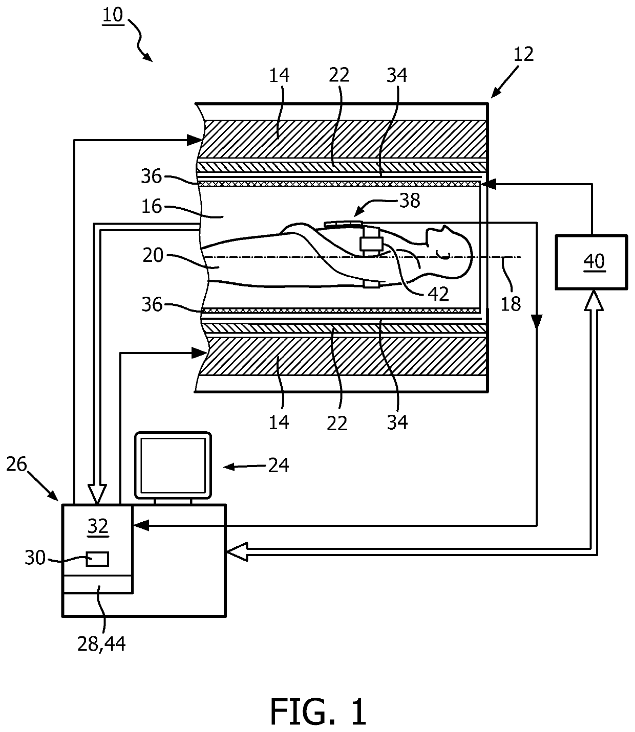
[0052]FIG. 1 shows a schematic illustration of a part of an embodiment of a magnetic resonance imaging system 10 configured for acquiring magnetic resonance images of at least a portion of a subject of interest 20, usually a patient. The magnetic resonance imaging system 10 comprises a scanning unit 12 having a main magnet 14. The main magnet 14 has a central bore that provides an examination space 16 around a center axis 18 for the subject of interest 20 to be positioned within, and is further provided for generating a static magnetic field B0 at least in the examination space 16. For clarity reasons, a customary table for supporting the subject of interest 20 has been omitted in FIG. 1. The static magnetic field B0 defines an axial direction of the examination space 16, aligned in parallel to the center axis 18. It is appreciated that the invention is also applicable to any other type of magnetic resonance imaging systems providing an examination region within a static magnetic fi...


PUM


Abstract

A method of operating a magnetic resonance imaging system (10) with regard to acquiring multiple-phase dynamic contrast-enhanced magnetic resonance images, the method comprising steps of acquiring (48) a first set of magnetic resonance image data (xpre) prior to administering a contrast agent to the subject of interest (20), by employing a water / fat magnetic resonance signal separation technique, determining (52) a first image of the spatial distribution of fat (Ipre) of at least the portion of the subject of interest (20), acquiring (50) at least a second set of magnetic resonance image data (x2) of at least the portion of the subject of interest (20) after administering the contrast agent to the subject of interest (20), by employing a water / fat magnetic resonance signal separation technique, determining (54) at least a second image of the spatial distribution of fat (I2ph) of at least the portion of the subject of interest (20), applying (56) an image registration method to the second image of the spatial distribution of fat (I2ph) with reference to the first image of the spatial distribution of fat (Ipre) for correcting a potential motion of the subject of interest (20); and a magnetic resonance imaging system (10) having a control unit (26) that is configured to carry out steps (56-64) of such a method; and a software module (44) for carrying out such a method, wherein the method steps (56-64) to be conducted are converted into a program code that is implementable in a memory unit (30) and is executable by a processor unit (32) of the magnetic resonance imaging system (10).

Description

CROSS REFERENCE TO RELATED APPLICATIONS[0001]This application is a U.S. national phase application of International Application No. PCT / IB2015 / 052354, filed on Mar. 31, 2015, which claims the benefit of U.S. provisional Application Ser. No. 61 / 980,668 filed on Apr. 17, 2014 and is incorporated herein by reference.FIELD OF THE INVENTION[0002]The invention pertains to a method of operating a magnetic resonance imaging system with regard to acquiring multiple-phase dynamic contrast-enhanced magnetic resonance images, and a magnetic resonance imaging system being operated by employing such a method.BACKGROUND OF THE INVENTION[0003]In the art of magnetic resonance imaging it is known to employ dynamic contrast-enhanced (DCE) imaging for obtaining physiological information like organ functions and perfusion, for instance by administering a contrast agent to a subject of interest, usually a patient, for tumor assessment. Several types of contrast agents and ways of administering are known ...

Claims


Application Information

Patent Timeline
Patent Type & Authority Patents(United States)
IPC IPC(8): A61B5/055A61B5/00G01R33/565G06T7/00G01R33/563G01R33/56G01R33/48
CPCA61B5/055A61B5/4869A61B5/7207G01R33/4828G01R33/56509G06T7/0012A61B5/4872G01R33/5601G01R33/56366G06T2207/10088G06T2207/30096
Inventor GDANIEC, NADINEBOERNERT, PETERDONEVA, MARIYA IVANOVAPEDROSA, IVAN
Owner BOARD OF RGT THE UNIV OF TEXAS SYST
Why Eureka
Try Eureka