Imaging method, controller and imaging system, for monitoring a patient post EVAR

a controller and imaging system technology, applied in the field of imaging methods, can solve the problems of increased risk of rupture, untreated, death, etc., and achieve the effect of convenient and less harmful approach

Active Publication Date: 2022-04-12
KONINKLJIJKE PHILIPS NV
View PDF5 Cites 0 Cited by
  • Summary
  • Abstract
  • Description
  • Claims
  • Application Information

AI Technical Summary

Benefits of technology

[0020]The invention thus provides a post-EVAR monitoring method in which two scans taken at different times can be compared to assess aneurysm evolution. The scans are compared by identifying the same recognizable features in the stent in the two images, aligning the images based on the recognizable features and then applying a registration process based on the recognizable features. By applying a registration approach based on stent matching, the operator can avoid bias between different measurements in time, avoid an invasive procedure and apply the method to different imaging modes (e.g. CT, MRI and US). The first 3D volume scan may be performed shortly after the stent graft procedure and may have been performed by a different clinician to the second 3D volume scan which is when the change in size of the aneurysm is to be analyzed.
[0026]The registration applied to the first and second scans may be a user-guided registration. The method may also include a further step after the registration of applying an imaged-based refinement of the registration. Both of these steps can be used to improve any sub-optimal alignment of the images.

Problems solved by technology

And as an aneurysm increases in size, the risk of rupture increases.
A ruptured aneurysm can lead to bleeding.
A ruptured AAA typically leads to hypovolemic shock and, if left untreated, death.
A type I endoleak is more serious, and results from the stent not sealing properly at the proximal or distal ends of the stent.
Less common but serious complications include type III and type IV endoleaks, which involve mechanical failure of the stent itself, which may result in surgical intervention.
Life-long surveillance after EVAR is troublesome for patients and physicians alike.
The surveillance requires significant resources, and may be associated with harm owing to the risk of potentially unnecessary interventions, repeated exposure to radiation, and contrast nephrotoxicity from the contrast agent.

Method used


Image

  • Imaging method, controller and imaging system, for monitoring a patient post EVAR
  • Imaging method, controller and imaging system, for monitoring a patient post EVAR
  • Imaging method, controller and imaging system, for monitoring a patient post EVAR

Examples


Embodiment Construction

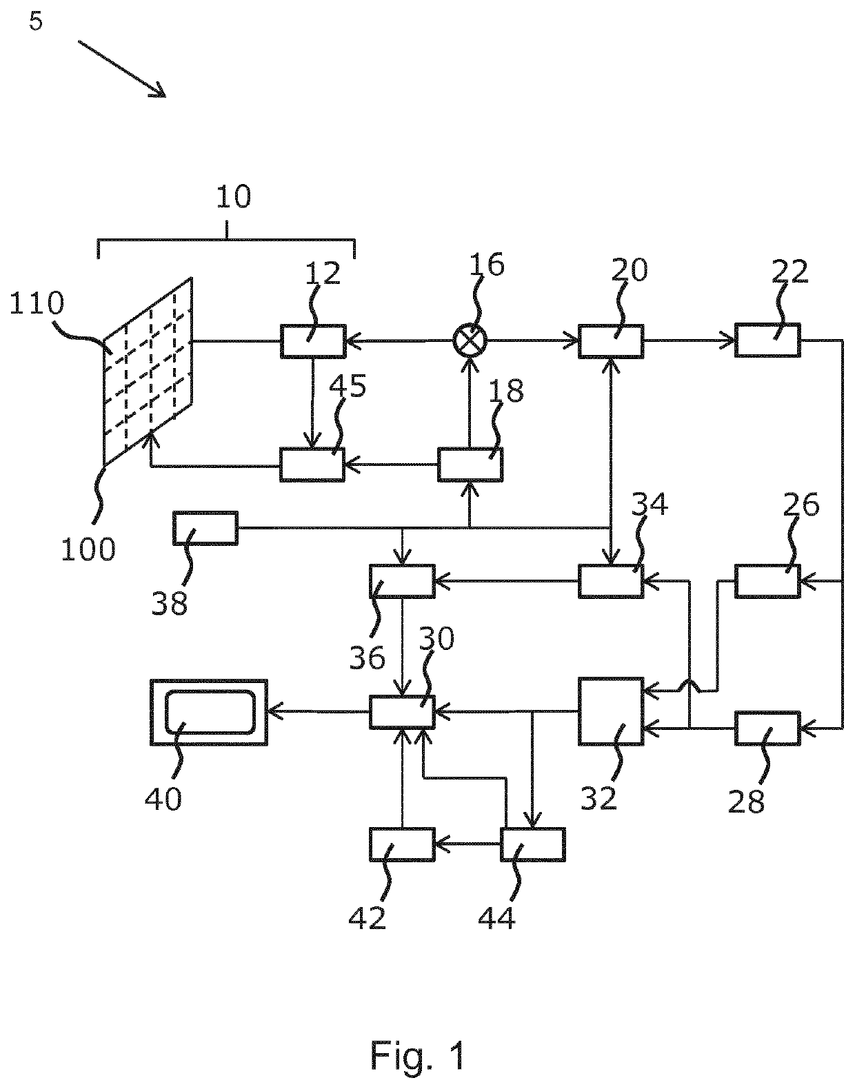
[0045]The invention provides a method of patient monitoring following EVAR using 3D-imaging to show any changes in size of the stent over time.

[0046]The invention provides a method of patient monitoring following endovascular aneurysm repair (EVAR) with a stent graft. It particularly applies to abdominal aortic aneurysm repair.

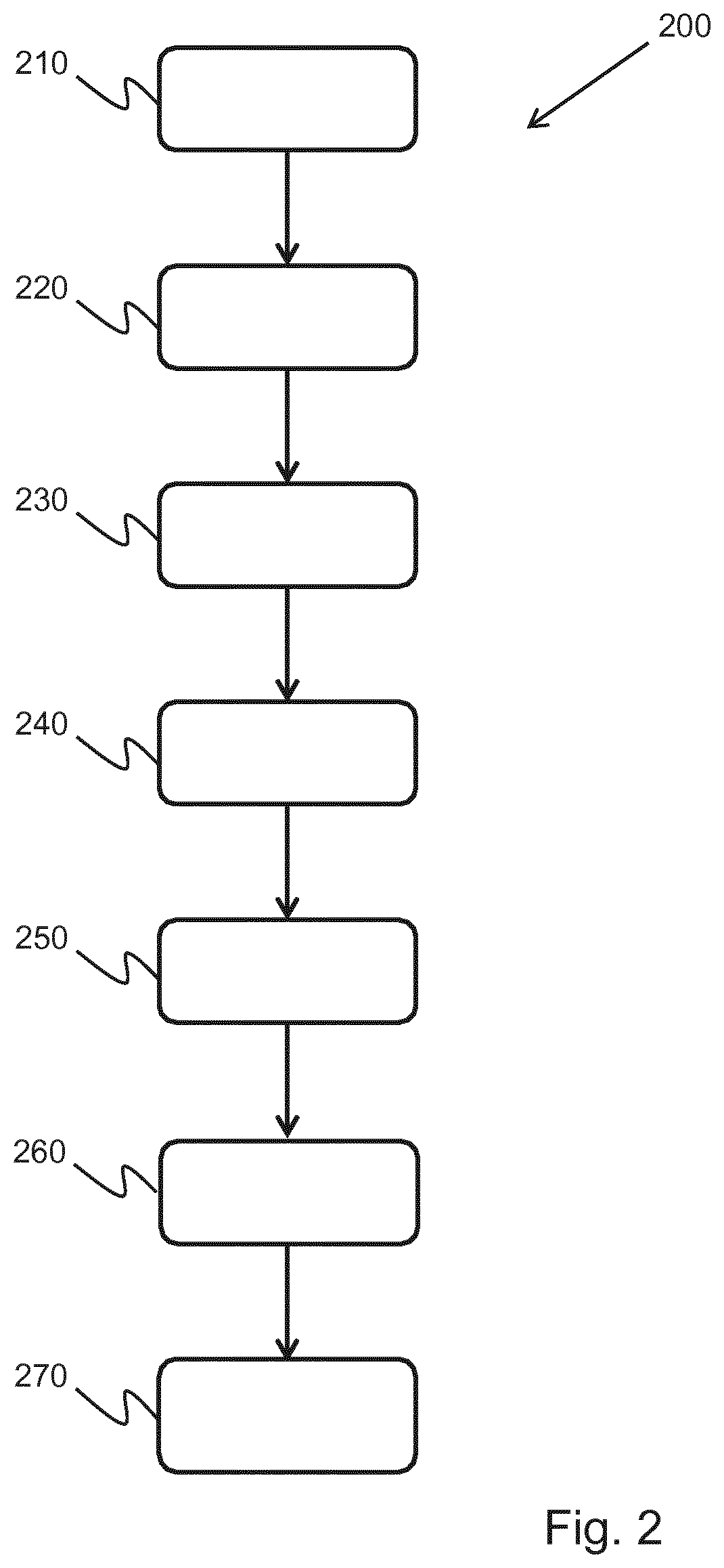
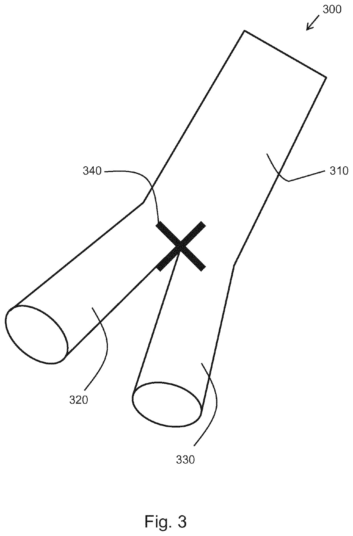
[0047]A first 3D volume scan of the stent in situ is provided and a second, subsequent 3D volume scan of the stent is also provided. One or more fiducial markers are generated in a first image derived from the first scan and in a second image derived from the second scan. The fiducial markers identify recognizable features of the stent, such as the bifurcation point in the stent graft. A rigid 3D transform mapping based on the one or more fiducial markers is extracted and then a registration is applied to the first and second scans based on the 3D rigid transform between the first and second images. A size value is derived at the same location in the aneurysm ...


PUM


Abstract

The invention provides a method of patient monitoring following endovascular aneurysm repair (EVAR) with a stent graft. It particularly applies to abdominal aortic aneurysm repair. A first 3D volume scan of the stent in situ is provided and a second, subsequent 3D volume scan of the stent is also provided. One or more fiducial markers are generated in a first image derived from the first scan and in a second image derived from the second scan. The fiducial markers identify recognizable features of the stent, such as the bifurcation point in the stent graft. A rigid 3D transform mapping based on the one or more fiducial markers is extracted and then a registration is applied to the first and second scans based on the 3D rigid transform between the first and second images. A size value is derived at the same location in the aneurysm from the first and second scans. The same location is determined with reference to the registered first and second images of the stent. A comparison of the size value from the first and second scans is used to assess a change in size of the aneurysm between the first and second scans. This change in size allows the clinician to evaluate disease progression.

Description

RELATED APPLICATIONS[0001]This application is the U.S. National Phase application under 35 U.S.C. § 371 of International Application No. PCT / EP2018 / 068889 (published as WO2019016057A), filed on Jul. 12, 2018, which claims priority to and the benefit of Provisional Application No. 62 / 533,308 (now U.S. Patent Application Publication No. 2020155102A1), filed Jul. 17, 2017 and of European Application No. 17183437.7 (published as EP3435382A1), filed Jul. 27, 2017. These applications are hereby incorporated by reference herein.FIELD OF THE INVENTION[0002]This invention relates to an imaging method and apparatus for monitoring a patient post-endovascular aneurysm repair (EVAR). It also provides a computer program and associated hardware to implement the imaging method.BACKGROUND OF THE INVENTION[0003]An aneurysm is a localized dilation of a blood vessel and is characterized by a bulge in the wall of the blood vessel at the point of the aneurysm. Aneurysms may occur in any blood vessel. The...

Claims


Application Information

Patent Timeline
Patent Type & Authority Patents(United States)
IPC IPC(8): A61B6/00G16H50/50G16H30/40A61B5/00A61B6/03A61B8/00
CPCA61B6/504A61B5/4842A61B6/032A61B6/463A61B6/5247A61B8/54G16H30/40G16H50/50
Inventor ROUET, LAURENCECOLLET-BILLON, ANTOINEDUFOUR, CECILEENTREKIN, ROBERT RANDALL
Owner KONINKLJIJKE PHILIPS NV
Why Eureka
Try Eureka