Enhancing ultrasound images

Active Publication Date: 2022-06-28
KONINKLJIJKE PHILIPS NV
View PDF24 Cites 1 Cited by
  • Summary
  • Abstract
  • Description
  • Claims
  • Application Information

AI Technical Summary

Benefits of technology

[0004]There may thus be a need to provide image data showing the current situation, for example in a region of interest of a patient, wherein the image data requires reduced and minimum interpretation and combination skill on the side of the user.
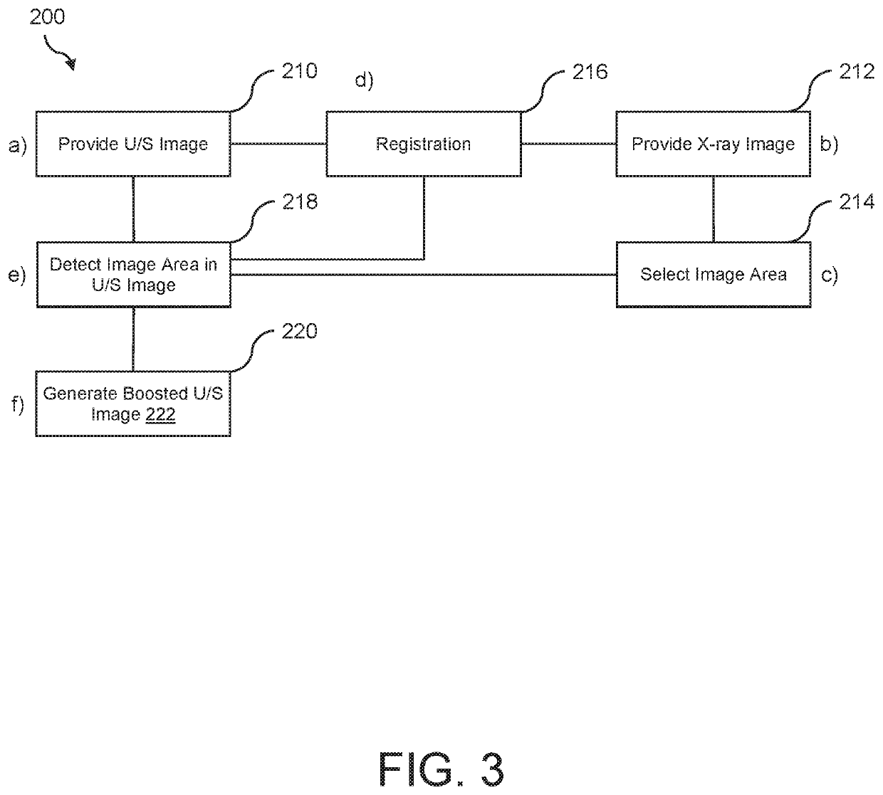
[0006]According to a first aspect of the present invention, an image processing for enhancing ultrasound images is provided, comprising an image data input unit, a central processing unit, and a display unit. The image data input unit is configured to provide an ultrasound image of a region of interest of an object, and to provide an X-ray image of the region of interest of the object. The central processing unit is configured to select a predetermined image area in the X-ray image. The central processing unit is further configured to register the ultrasound image and the X-ray image, and to detect the predetermined area in the ultrasound image based on the registered selected predetermined image area. The central processing unit is further configured to highlight at least a part of the detected area in the ultrasound image to generate a boosted ultrasound image. Thus, the term “boosted” or “boosting” in the invention refers to an improvement for a user in the visibility of an object. The display unit is configured to provide the boosted ultrasound image as guiding information on a display area.
[0010]By selecting the predetermined image area in the X-ray image, and by detecting the predetermined image area in the ultrasound image based on the registration, it is possible to enhance an ultrasound image based on information that is available in the X-ray image. Thus, image content, or image information, of the X-ray image is transferred to the ultrasound image. This provides image information that can be provided to the user, which image information is presented in one image, thus facilitating the viewing and understanding process of the provided image information.
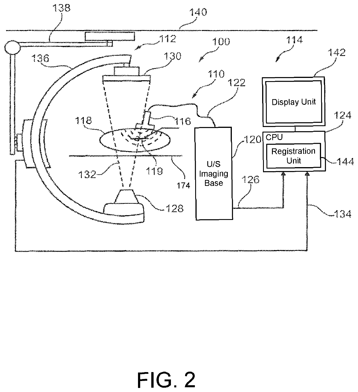
[0020]According to an aspect, the present invention proposes to boost an image by improving the visualization of ultrasound images or ultrasound 3D volumes. Such a boosting may be obtained by enhancing, for example colorizing, target objects, such as catheters, prosthesis, clips or the like, which can easily be detected and tracked in a fluoroscopy. Alternatively such a boosting may be obtained by outlining these target objects. As mentioned above, for example in case of endovascular interventions, X-ray images, such as fluoroscopic images allow a good visualization of endovascular tools, but it does not allow a visualization of soft tissue (without the use of contrast agent) or a 3D visualization. Ultrasound allows a 2D or 3D visualization including soft tissues. However the contrast between tools and soft tissues is very low. Further, in two-dimensional ultrasound images, only a part of a tool may be visible. Therefore the interpretation of such image is not straight forward. The present invention proposes to perform the detection, or the selection, of target objects, or other predetermined image area in fluoroscopic images, in which they are properly contrasted. Further, the target objects may be tracked in the X-ray images. Based on a registration, for example a live registration, of ultrasound images and X-ray images, the target objects, or predetermined image areas can be detected in the ultrasound image, and the target objects can be enhanced in the ultrasound images, for example by colorizing them. It must be noted that the X-ray image data is only used to detect in the ultrasound images target objects or predetermined image area whose detection could be very challenging otherwise. Indeed the high contrast of interventional tools in X-ray images makes easier their detection and tracking. Even if in ultrasound images they are poorly contrasted (with respect to soft tissues), their detection is facilitated by the position and shape priors that arise from the X-Ray detection and that are based on the registration of the two image modalities. Instead of combining the two images, the transfer from the X-ray image to the ultrasound image is provided for detection and / or positioning purposes only. However, the transfer of the positioning data from the X-ray image to the ultrasound image is adding further information to the ultrasound image, which information can not be retrieved from the original ultrasound image itself.

Problems solved by technology

However, it has also been shown that providing fluoroscopic images and ultrasound images next to each other requires attention and awareness of the user mentally combining the two different contents shown in the respective images.

Method used


Image

  • Enhancing ultrasound images
  • Enhancing ultrasound images
  • Enhancing ultrasound images

Examples


Embodiment Construction

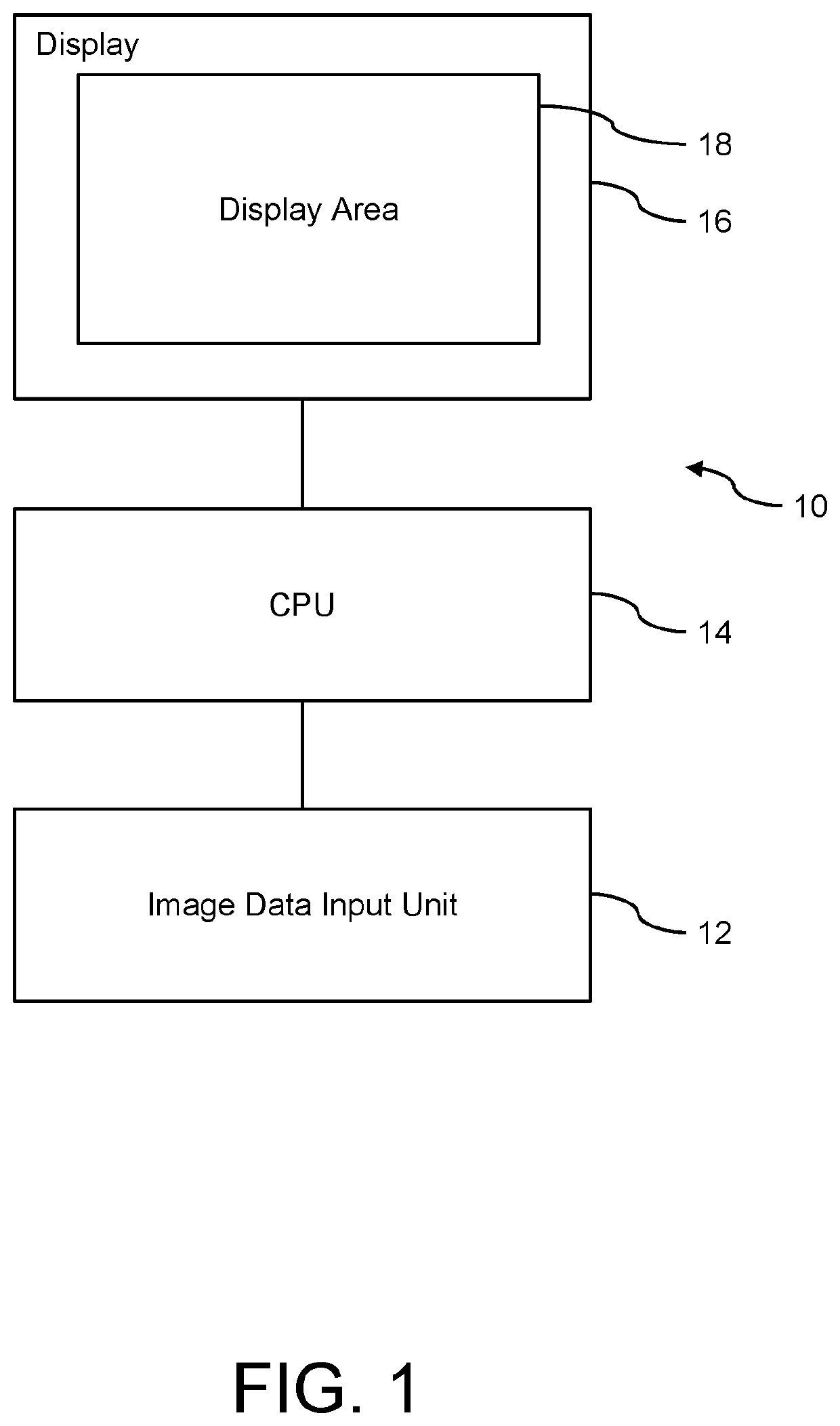
[0030]FIG. 1 shows an image processing device 10 for enhancing ultrasound images, comprising an image data input unit 12, a central processing unit 14, and a display unit 16. The image data input unit 12 is configured to provide an ultrasound image of a region of interest of an object, and to provide an X-ray image of the region of interest of the object.

[0031]The central processing unit 14 is configured to select a predetermined image area in the X-ray image, to register the ultrasound image and the X-ray image, to detect the predetermined area in the ultrasound image based on the registered selected predetermined image area, and to highlight at least a part of the detected area in the ultrasound image to generate a boosted ultrasound image. The display unit is configured to provide the boosted ultrasound image as guiding information on a display area 18.

[0032]In a further example (not further shown), the central processing unit is configured to project the selected area into the u...


PUM


Abstract

The present invention relates to image processing for enhancing ultrasound images. In order to provide image data showing the current situation, for example in a region of interest of a patient, an image processing device (10) for enhancing ultrasound images is provided that comprises an image data input unit (12), a central processing unit (14), and a display unit (16). The image data input unit is configured to provide an ultrasound image of a region of interest of an object, and to provide an X-ray image of the region of interest of the object. The central processing unit is configured to select a predetermined image area in the X-ray image, to register the ultrasound image and the X-ray image, to detect the predetermined area in the ultrasound image based on the registered selected predetermined image area, and to highlight at least a part of the detected area in the ultrasound image to generate a boosted ultrasound image. The display unit is configured to provide the boosted ultrasound image as guiding information on a display area (18).

Description

CROSS-REFERENCE TO PRIOR APPLICATIONS[0001]This application is the U.S. National Phase application under 35 U.S.C. § 371 of International Application Serial No. PCT / IB2013 / 059793, filed on Oct. 30, 2013, which claims the benefit of EP Application No. 12306368.7, filed on Nov. 6, 2012. These applications are hereby incorporated by reference herein.FIELD OF THE INVENTION[0002]The present invention relates to an image processing device for enhancing ultrasound images, to a medical imaging system, and to a method for enhancing ultrasound images, as well as to a computer program element and to a computer-readable medium.BACKGROUND OF THE INVENTION[0003]In medical examinations, such as endovascular interventions, different types of medical imaging can be used, for example the positioning of a mitral clip is performed under fluoroscopic X-ray imaging and ultrasound imaging for guidance purposes. The X-ray images are used for visualizing tools, such as catheters, clips, or the like, and als...

Claims


Application Information

Patent Timeline
Patent Type & Authority Patents(United States)
IPC IPC(8): A61B6/00A61B6/12A61B8/00G06T15/08G06T7/30G06T7/62G06T7/70A61B8/08G06T1/00
CPCG06T15/08A61B6/12A61B6/4417A61B6/469A61B6/5247A61B8/0833A61B8/0841A61B8/4416A61B8/461A61B8/463A61B8/469A61B8/5261G06T1/0007G06T7/30G06T7/62G06T7/70A61B6/4441A61B6/487A61B8/466G06T2207/10121G06T2207/10132G06T2207/20221G06T2207/30004G06T2215/12
Inventor NEMPONT, OLIVIER PIERRECATHIER, PASCAL YVES FRANCOISFLORENT, RAOUL
Owner KONINKLJIJKE PHILIPS NV
Why Eureka
Try Eureka