System of joint brain tumor and cortex reconstruction

a brain tumor and cortex technology, applied in the field of joint brain tumor and cortex reconstruction, can solve the problems of system failure to take into account deformed anatomy which arises, high labor intensity of manual processing, and complex and difficult process of analyzing magnetic resonance image data for the detection, reconstruction and imaging of tumors

Active Publication Date: 2022-07-19
SONY CORP
View PDF14 Cites 0 Cited by
  • Summary
  • Abstract
  • Description
  • Claims
  • Application Information

AI Technical Summary

Benefits of technology

[0010]A fully automatic brain tumor and tumor-aware cortex reconstruction system to improve the healthcare experience with machine intelligence. The disclosed technology receives multi-modal MRI data (T1, T1c, T2, T2-Flair) performs joint reconstruction and visualization of tumors, white matter, and gray matter surfaces automatically.
[0012]The disclosed technology has been validated by a publicly-available challenge dataset, specifically BraTS2015. A qualitative evaluation indicated that the disclosed method can successfully reconstruct both tumor and cortical surfaces for all relevant datasets.

Problems solved by technology

As manual processing is so labor intensive, automated approaches are being sought.
Yet, analyzing magnetic resonance image (MRI) data for the detection, reconstruction and imaging of tumors is a complex and difficult process.
For example, systems typically fail to take into account deformed anatomy which arises in response to a tumor.
In addition, many automated approaches can distort the reconstruction, such as by overcompensation.
Furthermore, the use of available low resolution datasets (e.g., BraTS2105) limits discernment and imaging capabilities.
However, it was very problematic to find correct (useful) seeds in the low resolution patient data, and often proper areas were scattered or their positioning represented false connections to other areas.

Method used


Image

  • System of joint brain tumor and cortex reconstruction
  • System of joint brain tumor and cortex reconstruction
  • System of joint brain tumor and cortex reconstruction

Examples


Embodiment Construction

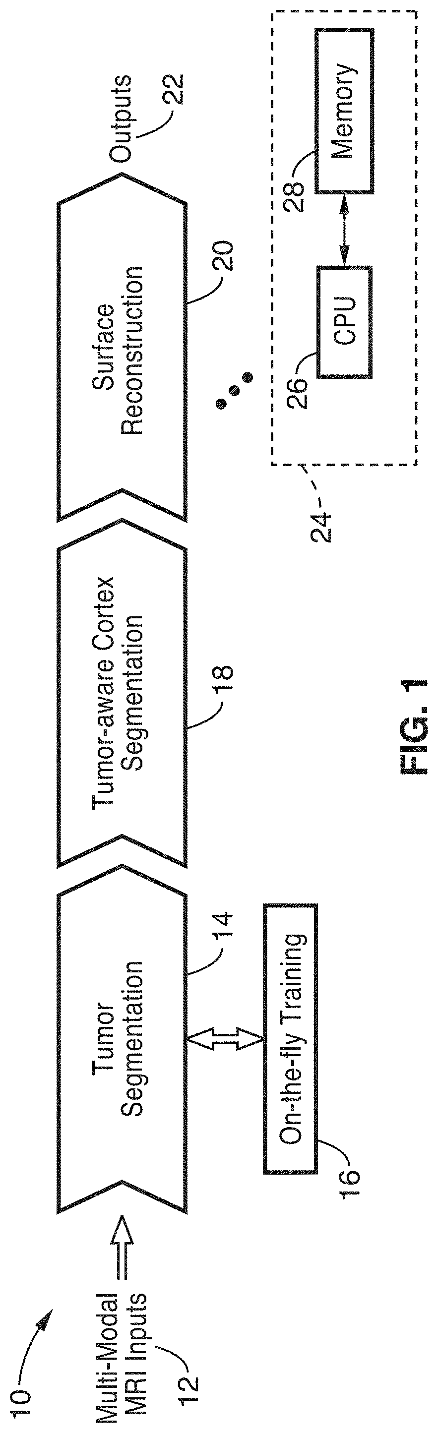
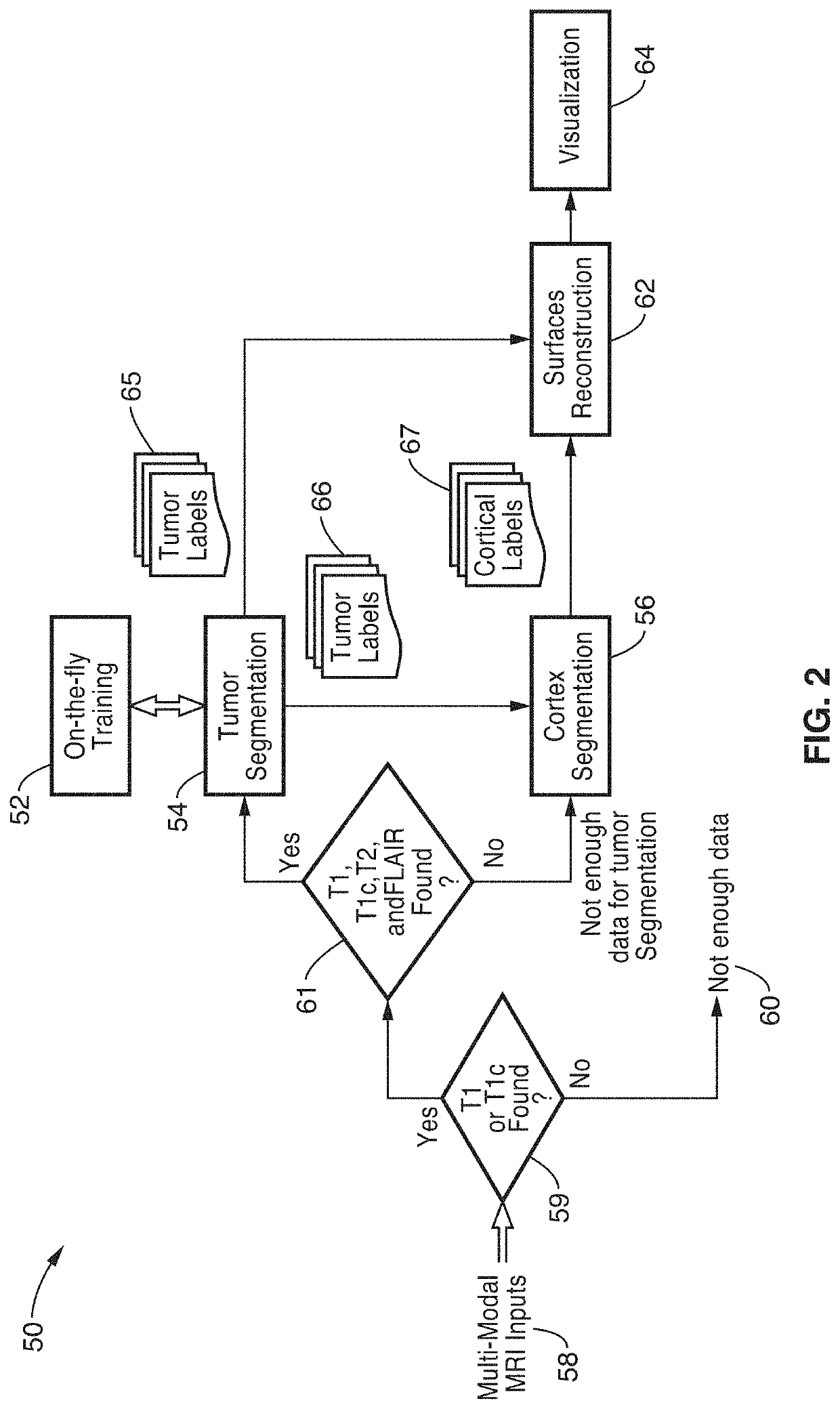
[0026]FIG. 1 illustrates an overview 10 of joint brain tumor and cortex reconstruction of the present disclosure. Multi-modal inputs (e.g., T1, T1c, T2, and FLAIR) are received 12 from a magnetic resonance imaging (MRI) system, for processing according to the present disclosure. This data is received into tumor segmentation module 14 which is configured for performing on-the-fly training 16. Outputs from tumor segmentation are received in a module 18 for performing tumor-aware cortex segmentation. Outputs from tumor-aware cortex segmentation are received in module 20 for performing surface reconstructions, before generating an output 22 which delineates white matter, grey matter, tumor edema, and a tumor active core.

[0027]Processing 24 within each of these modules is shown exemplified with at least one computer (CPU) 26 along with memory 28. It will be appreciated that instructions / programming stored on memory (computer readable media) 28 is executable on computer processor 26. The ...


PUM


Abstract

System for performing fully automatic brain tumor and tumor-aware cortex reconstructions upon receiving multi-modal MRI data (T1, T1c, T2, T2-Flair). The system outputs imaging which delineates distinctions between tumors (including tumor edema, and tumor active core), from white matter and gray matter surfaces. In cases where existing MRI model data is insufficient then the model is trained on-the-fly for tumor segmentation and classification. A tumor-aware cortex segmentation that is adaptive to the presence of the tumor is performed using labels, from which the system reconstructs and visualizes both tumor and cortical surfaces for diagnostic and surgical guidance. The technology has been validated using a publicly-available challenge dataset.

Description

CROSS-REFERENCE TO RELATED APPLICATIONS[0001]Not ApplicableSTATEMENT REGARDING FEDERALLY SPONSORED RESEARCH OR DEVELOPMENT[0002]Not ApplicableINCORPORATION-BY-REFERENCE OF COMPUTER PROGRAM APPENDIX[0003]Not ApplicableNOTICE OF MATERIAL SUBJECT TO COPYRIGHT PROTECTION[0004]A portion of the material in this patent document is subject to copyright protection under the copyright laws of the United States and of other countries. The owner of the copyright rights has no objection to the facsimile reproduction by anyone of the patent document or the patent disclosure, as it appears in the United States Patent and Trademark Office publicly available file or records, but otherwise reserves all copyright rights whatsoever. The copyright owner does not hereby waive any of its rights to have this patent document maintained in secrecy, including without limitation its rights pursuant to 37 C.F.R. § 1.14.BACKGROUND1. Technical Field[0005]The technology of this disclosure pertains generally to ana...

Claims


Application Information

Patent Timeline
Patent Type & Authority Patents(United States)
IPC IPC(8): A61B5/00A61B5/055G06T7/00G06K9/62G01R33/56G06T7/11G06T7/162G01R33/50G06V10/776
CPCA61B5/4064A61B5/0035A61B5/0042A61B5/055A61B5/7267G01R33/5608G06K9/6262G06K9/6282G06T7/0014G06T7/11G06T7/162A61B2576/026G01R33/50G06T2207/10088G06T2207/20081G06T2207/20112G06T2207/30016G06T2207/30096G06V2201/03G16H30/40G16H50/70G06V10/776G06F18/24323G06F18/217
Inventor CHOU, CHEN-RUISONG, BILIU, MING-CHANG
Owner SONY CORP
Why Eureka
Try Eureka