P63 inactivation for the treatment of heart failure

a heart failure and inactivation technology, applied in the field of molecular biology, cell biology, cell therapy, medicine, can solve the problem of low reprogramming efficiency

Active Publication Date: 2022-08-23
BAYLOR COLLEGE OF MEDICINE
View PDF12 Cites 0 Cited by
  • Summary
  • Abstract
  • Description
  • Claims
  • Application Information

AI Technical Summary

Benefits of technology

[0008]In particular embodiments of the disclosure, p63, p53, and / or p21 inactivation increases the transdifferentiation efficiency of cardiac cells (such as fibroblasts) into cardiomyocytes. As described herein, the partial or complete inactivation of the p63, p53, and / or p21 genes is a novel therapeutic intervention that allows the reprogramming of fibroblasts (for example) into cardiomyocytes at a much higher efficiency. In vitro studies confirm that 45% of p63 knockout mouse embryonic fibroblasts express cardiac Troponin T (cTnT), which is a highly specific marker for the cardiomyocyte lineage. p63 inactivation through shRNA or siRNA (for example) is a unique intervention that is a clinically relevant therapy for the treatment of any cardiac medical condition, including heart failure.

Problems solved by technology

Nevertheless, the major obstacle in the implementation of this therapy is the low efficiency of reprogramming.

Method used


Image

  • P63 inactivation for the treatment of heart failure
  • P63 inactivation for the treatment of heart failure
  • P63 inactivation for the treatment of heart failure

Examples


example 1

P63 Inactivation Increases the Transdifferentiation Efficiency of Fibroblasts into Cardiomyocytes

[0294]Cellular regenerative therapy has emerged as a treatment for a number of degenerative diseases, most notably ischemic heart disease, which is the number one cause of death in the world. Several strategies have been proposed, including transdifferentiation and the formation of induced pluripotent stem cells. Transdifferentiation has been attempted with transcription factors such as Gata4, Mef2c and Tbx5 (GMT), while iPS cells may be formed using the Yamanaka factors (Oct4, Sox2, Klf4, c-myc) or knocking down genes involved in regulating the cell cycle, such as p53.

[0295]While it has been reported that murine cells that are p63 deficient display a high degree of plasticity, no distinct strategy has been developed for their application in ischemic heart disease. Specifically, it has not been described how the p63 deficient cells may be “driven” down the cardiomyocyte lineage. The atta...

example 2

[0299]P63 Inactivation and Addition of Hand2, Myocardin Increases Formation of CTNT+ Cells from Murine Embryonic Fibroblasts

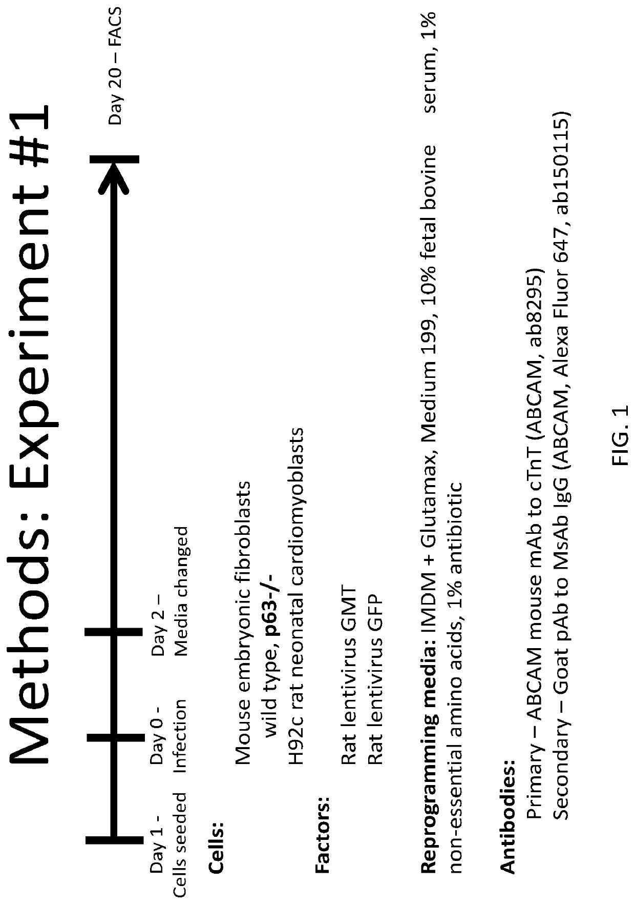
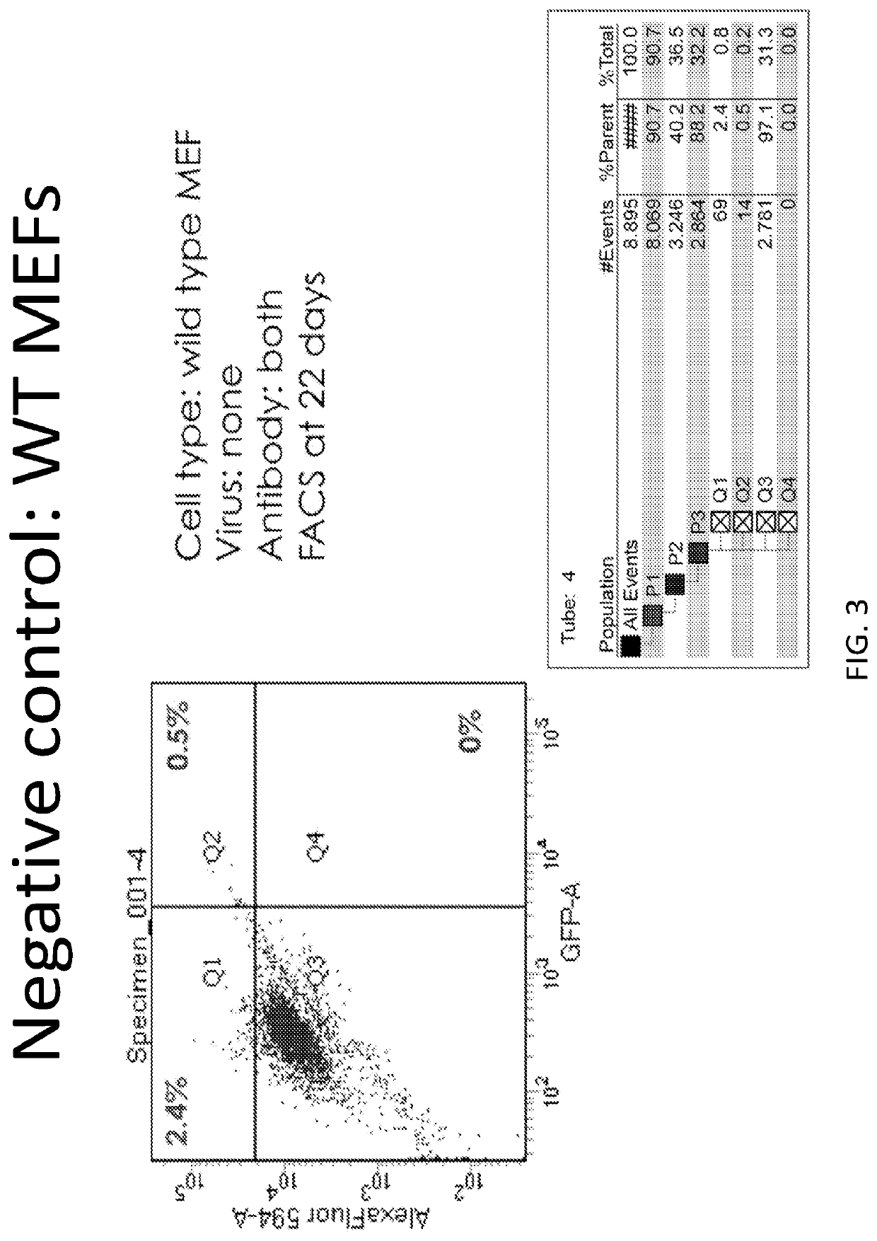
[0300]FIG. 12 illustrates an experimental design for analyzing transdifferentiation of fibroblasts into cardiomyocytes in vitro. FIG. 13 shows FACS data for unstained and stained wild-type MEFs (negative controls). FIG. 14 shows wild type MEFs treated with lentiviral GFP and lentiviral GMT. In FIG. 15, wild-type MEFs were given the noted factors (lentiviral Hand2 & Myocardin) and exposed to both antibodies. In FIG. 16, shows a positive control, h9c2 neonatal rat cardiomyoblasts. FIG. 17 shows the p63 − / − (which concerns the inactivation of both the TA and Delta isoforms of the p63 gene) MEFs where no factors were provided. The left side shows the control stained with only the secondary antibody while the right shows cells stained with both antibodies. FIG. 18 demonstrates the double KO p63− / − MEFs where GFP (left side shows staining only with secondary as a con...

example 3

P63 Inactivation and the Addition of Several Other Factors May Further Enhance the Transdifferentiation of Fibroblasts into Cardiomyocytes

[0303]Examples 1 and 2 illustrated two embodiments of applications of this disclosure. However, there are several other transcription factors that may be used to further enhance the efficiency: Gata4, Mef2c, Tbx5, miR-133, miR-1, Oct4, Klf4, c-myc, Sox2, Mesp1, Brachyury, Nkx2.5, ETS2, ESRRG, Mrtf-A, MyoD, ZFPM2 (in nucleic acid or polypeptide or peptide form, in specific embodiments). Adjunctive therapy could comprise ITD-1 (TGF beta inhibitor), VEGF, surgery (coronary artery bypass), PCI, or medial therapy, for example.


PUM

PropertyMeasurementUnit
temperatureaaaaaaaaaa
ionic strengthaaaaaaaaaa
temperatureaaaaaaaaaa

Abstract

Embodiments of the disclosure include methods and compositions for in situ cardiac cell regeneration, including transdifferentiation of cardiac cells to cardiomyocytes. In particular embodiments, in situ cardiac cell regeneration encompasses delivery of p63 shRNA and one or both of Hand2 and myocardin, and in specific embodiments further includes one or more of Gata4, Mef2c, and Tbx5. In specific aspects of the disclosure, adult cardiac fibroblasts are reprogrammed into cardiomyocytes using viral vectors that harbor p63 shRNA and one or both of the transcription factors Hand2 and myocardin.

Description

[0001]This application is a national phase application under 35 U.S.C. § 371 that claims priority to International Application No. PCT / US2016 / 018735 filed Feb. 19, 2016 which claims the benefit of priority to U.S. Provisional Patent Application Ser. No. 62 / 118,573, filed Feb. 20, 2015, all of which are incorporated by reference herein in their entirety.STATEMENT REGARDING FEDERALLY SPONSORED RESEARCH OR DEVELOPMENT[0002]This invention was made with government support under HL121294 awarded by the National Institutes of Health. The government has certain rights in the invention.TECHNICAL FIELD[0003]Embodiments of the disclosure include at least the fields of molecular biology, cell biology, cell therapy, and medicine, including cardiac medicine.BACKGROUND OF THE INVENTION[0004]Despite medical and surgical innovations, heart disease remains the number one cause of death in the world. Given the poor regenerative capacity of the heart following myocardial infarction and the irreversible...

Claims


Application Information

Patent Timeline
Patent Type & Authority Patents(United States)
IPC IPC(8): C12N15/113A61K38/17A61K31/47C07K14/47C07K14/82C12N5/077A61K9/00A61K31/713A61K48/00C12N15/86
CPCC12N15/113A61K9/0019A61K31/47A61K31/713A61K38/1709A61K48/0058C07K14/4702C07K14/4746C07K14/82C12N5/0657C12N15/1135C12N15/86C12N2310/14C12N2310/531C12N2320/31C12N2501/60C12N2710/10043C12N2740/15043
Inventor PATEL, VIVEKKUMAR B.WANG, HONGRANSINGH, VIVEK P.REINEKE, ERIN LYNNMATHISON, MEGUMICOONEY, AUSTIN J.ROSENGART, TODD
Owner BAYLOR COLLEGE OF MEDICINE
Why Eureka
Try Eureka