Method and apparatus for non-invasive blood constituent monitoring

a non-invasive and blood constituent technology, applied in the field of non-invasive blood constituent monitoring, can solve the problems of failure to isolate and resolve individual and specific scattering and absorption coefficients of desired constituents

Inactive Publication Date: 2001-11-08
HEMA METRICS
View PDF0 Cites 126 Cited by
  • Summary
  • Abstract
  • Description
  • Claims
  • Application Information

AI Technical Summary

Benefits of technology

[0008] Thus, it is an object of the present invention to provide an improvement in the systems and methods for the non-invasive (transcutaneous) and continuous determination of the blood Hematocrit in living tissue.
[0009] It is yet another object of the present invention to provide an improvement in the systems and methods for the non-invasive (transcutaneous) and continuous determination of the blood constituents, including glucose, bilirubin, cholesterol, tissue water, etc. in living tissue.
[0011] It is yet another object of the present invention to provide a repeatable and reliable method and apparatus for the non-invasive determination of hematocrit transcutaneously and in real time even under varying physiological conditions.
[0033] In the present disclosure, FIG. 1 shows the finger 7 of an individual placed into a clam-shell type fixture 6 wherein the optical and other physical measurements can be easily performed. The clam-shell type holder allows for adaptability to various finger sizes. However, other fixture methods such as FIGS. 1B through 1E, can be used to obtain similar physical data as using the clam-shell fixture.THEORETICAL BASIS OF THE SPECTROPHOTOMETRIC AND MATHEMATICAL ANALYSIS FOR TRANSCUTANEOUS HEMATOCRIT MEASUREMENT
[0036] Earlier spectrophotometric techniques have fallen short of being able to fully characterize the individual blood absorbance coefficients. The following discussion demonstrates the method of decoupling, or isolating from the bulk tissue attenuation parameters (including the convoluted absorptive and scattering parameters) the individual blood absorptive constants. This unique method identifies, isolates and compartmentalizes each of the contributing biologic elements of the tissue media. This decoupling process can either isolate the blood absorbance of interest and / or eliminate the scattering contribution from the bulk media measurement.
[0133] If hematocrit is constant over a given time interval, averaging can eliminate system noise whose frequency components have corresponding periods much shorter than the interval. In addition, by observing the data variance during the interval it may be determined that the data is invalid. In the present system, the data acquisition rate is approximately 1000 data samples per second. This means that within a typical human pulse about 1000 samples of data are available for appropriate numerical analysis, averaging and qualification. Recognizing that both the intensity of light and the pressure in the transducer system are changing in time during the influx of blood is of great importance. Since the parametric relationship of .differential..alpha. / .d-ifferential.t as a function of .differential.P / .differential.t (where P is pressure) during the cardiac cycle should be linear, a multiplicity of data points facilitate qualification of the signal for accuracy and linearity. Whereas, prior techniques involving only the peak and valley values of the cardiac cycle require numerous pulses to qualify the data set. See FIGS. 8, 9 and 10.

Problems solved by technology

Even though the various patents indicate the need to utilize multiple wavelengths measured at different detection sites and / or the need to perform differential or ratiometric operations on the detected optical signal, all fail to isolate and resolve the individual and specific scattering and absorption coefficients of the desired constituent.
Secondly, the actual absorptive coefficient of the blood must then be decoupled or isolated from its proration factor as well.

Method used


Image

  • Method and apparatus for non-invasive blood constituent monitoring
  • Method and apparatus for non-invasive blood constituent monitoring
  • Method and apparatus for non-invasive blood constituent monitoring

Examples


Embodiment Construction

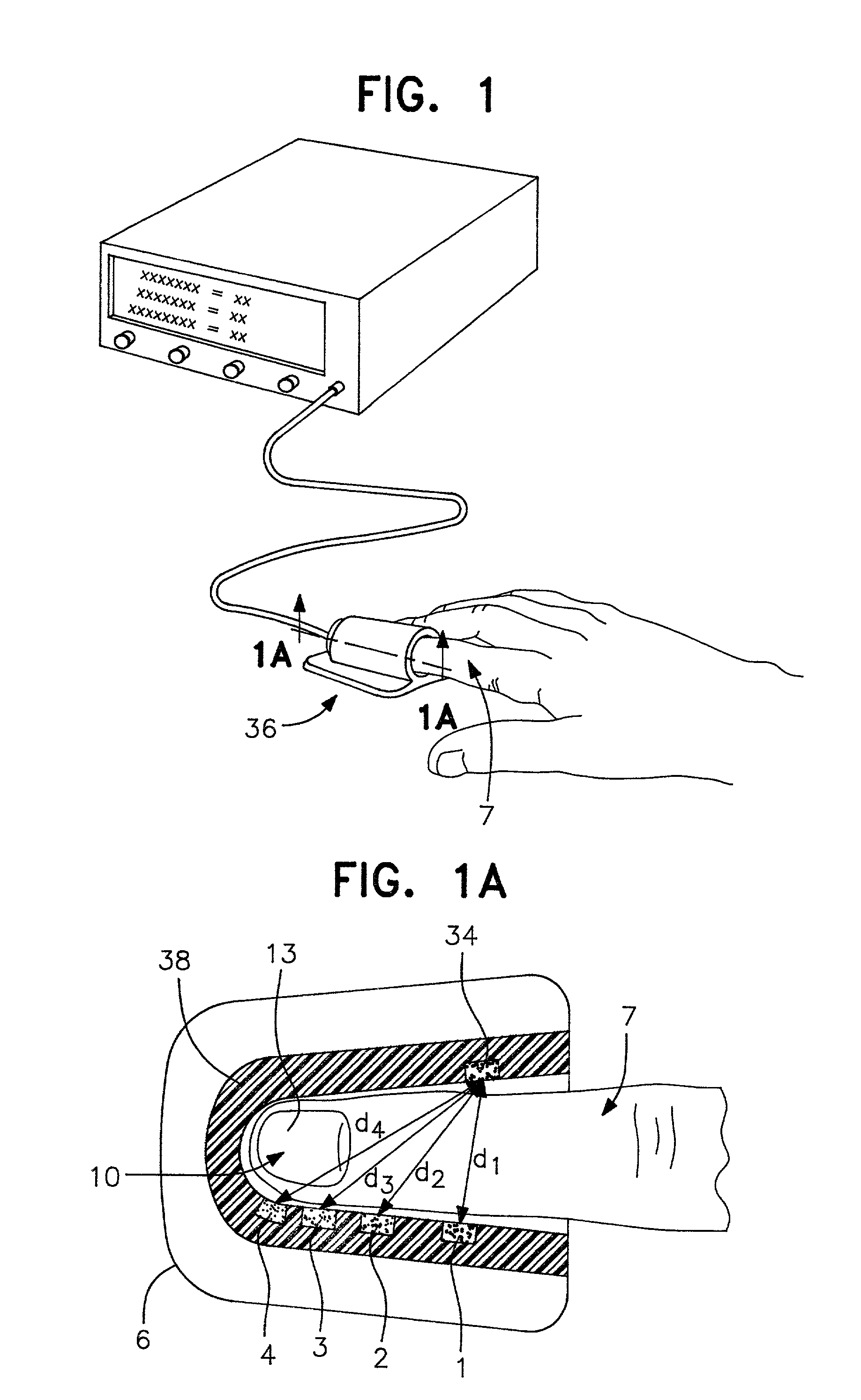
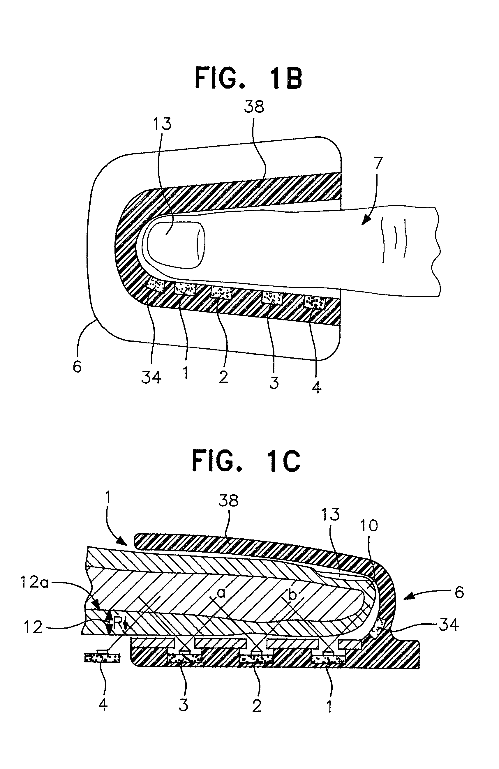
[0157] Physical embodiments as shown in FIG. 1 include the optical array, pressure transducer / balloon system and clam-shell fixture. Requisites of the preferred embodiment include a holder for the finger (or other tissue) such as seen in FIGS. 1 and 1A and 1B. This clam-shell fixture not only secures the tissue but also the optical array, and transducer system.

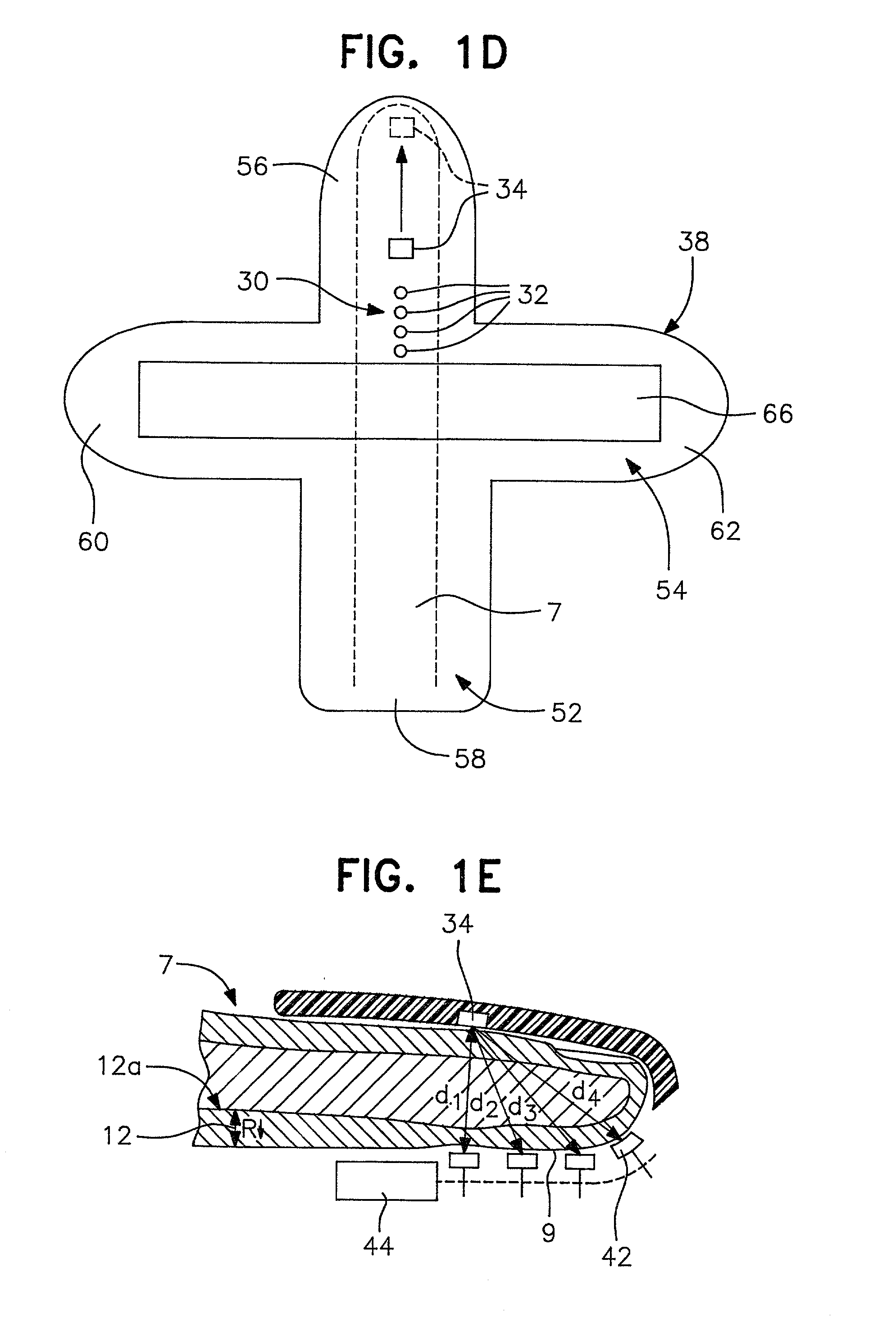
[0158] FIG. 1D is a schematic diagram for a mylar base member 38 that is shaped generally like a cross. As oriented in FIG. 1D, vertically extending portion 52 crosses with a horizontally extending portion 54 to yield top leg 56, bottom leg 58, and side legs 60, 62. In use, a finger 7 lies along the longitudinally extending portion 52 with the finger tip placed on the top leg 56 to properly cover the arrangement of LED's 32 and photodetector 34, which are arranged like those on FIGS. 1A-1C. A piezoelectric pressure transducer or strain gage 66 spans the horizontally extending portion 54 from near the tip of side leg 60 to the ...


PUM


Abstract

A system for determining a biologic constituent including hematocrit transcutaneously, noninvasively and continuously. A finger clip assembly includes including at least a pair of emitters and a photodiode in appropriate alignment to enable operation in either a transmissive mode or a reflectance mode. At least one predetermined wavelength of light is passed onto or through body tissues such as a finger, earlobe, or scalp, etc. and attenuation of light at that wavelength is detected. Likewise, the change in blood flow is determined by various techniques including optical, pressure, piezo and strain gage methods. Mathematical manipulation of the detected values compensates for the effects of body tissue and fluid and determines the hematocrit value. If an additional wavelength of light is used which attenuates light substantially differently by oxyhemoglobin and reduced hemoglobin, then the blood oxygen saturation value, independent of hematocrit may be determined. Further, if an additional wavelength of light is used which greatly attenuates light due to bilirubin (440 nm) or glucose (1060 nm), then the bilirubin or glucose value may also be determined. Also how to determine the hematocrit with a two step DC analysis technique is provided. Then a pulse wave is not required, so this method may be utilized in states of low blood pressure or low blood flow.

Description

[0001] The present invention is related to U.S. Pat. Nos. 5,372,136 and 5,499,627 the text and drawings of which are incorporated herein by reference as if reproduced in full.[0002] 1. The Field of the Invention[0003] The present invention relates to improvements in the systems and methods for non-invasively measuring one or more biologic constituent concentration values. More particularly, the present invention relates to non-invasive spectrophotometric systems and methods for quantitatively and continuously monitoring the hematocrit and other blood parameters.[0004] 2. The Prior Art[0005] Modern medical practice utilizes a number of procedures and indicators to assess a patient's condition. One of these indicators is the patient's hematocrit. Hematocrit (often abbreviated as HCT) is the volume expressed as a percentage of the patient's blood which is occupied by red corpuscles, commonly referred to as red blood cells. The present invention is presented in the context of hematocrit...

Claims


Application Information

Patent Timeline
Patent Type & Authority Applications(United States)
IPC IPC(8): A61B5/145A61B5/00A61B5/1455
CPCA61B5/14532A61B5/14535A61B5/14552A61B5/6826A61B5/6838A61B5/6843
Inventor STEUER, ROBERT R.MILLER, DAVID R.
Owner HEMA METRICS
Why Eureka
Try Eureka