Method and apparatus for endoscope system

a technology of endoscopy and endoscope, which is applied in the field of medical devices, can solve the problems of system failure to guide a physician to targeted areas, and the technique is problematic, and achieves the effect of improving the accuracy of endoscopy results

Inactive Publication Date: 2003-03-20
LACHOWICZ THEODORE COLLATERAL AGENT
View PDF4 Cites 39 Cited by
  • Summary
  • Abstract
  • Description
  • Claims
  • Application Information

AI Technical Summary

Problems solved by technology

These techniques, however, have been problematic in the past.
This technique can be disadvantageous, because the guidewire guidance into body tissue requires arbitrary trial and error guidance and the system does not allow a doctor to navigate the guidewire to targeted cells by using images from the endoscope.
Both of these systems lack the capability to guide a physician to targeted areas without having the physician at least make some educated guesses as to where that targeted area may be.

Method used


Image

  • Method and apparatus for endoscope system
  • Method and apparatus for endoscope system
  • Method and apparatus for endoscope system

Examples


Embodiment Construction

[0041] Although the present invention is described below in connection with specific embodiments, it will be appreciated that the invention is not limited to the described embodiments.

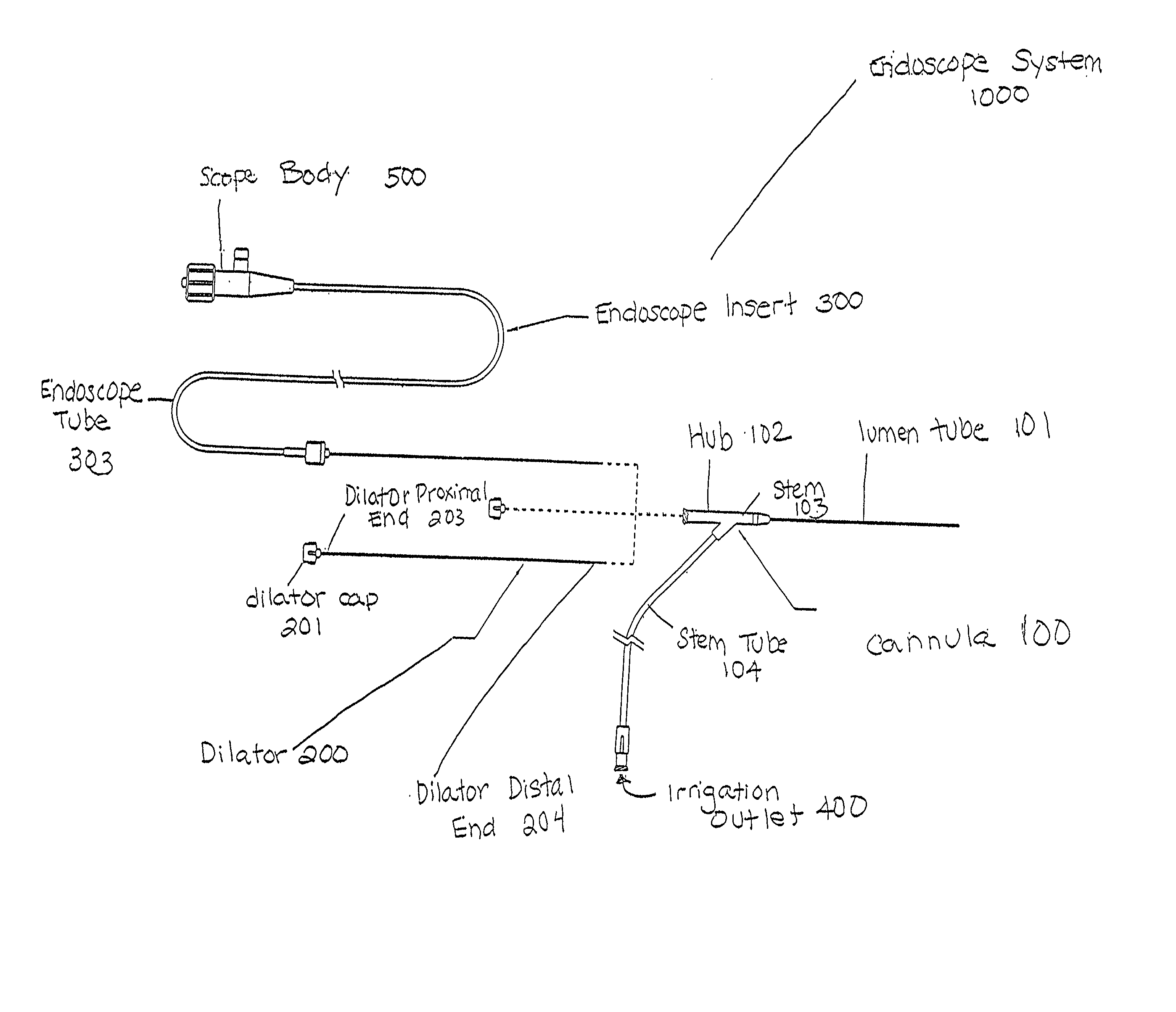
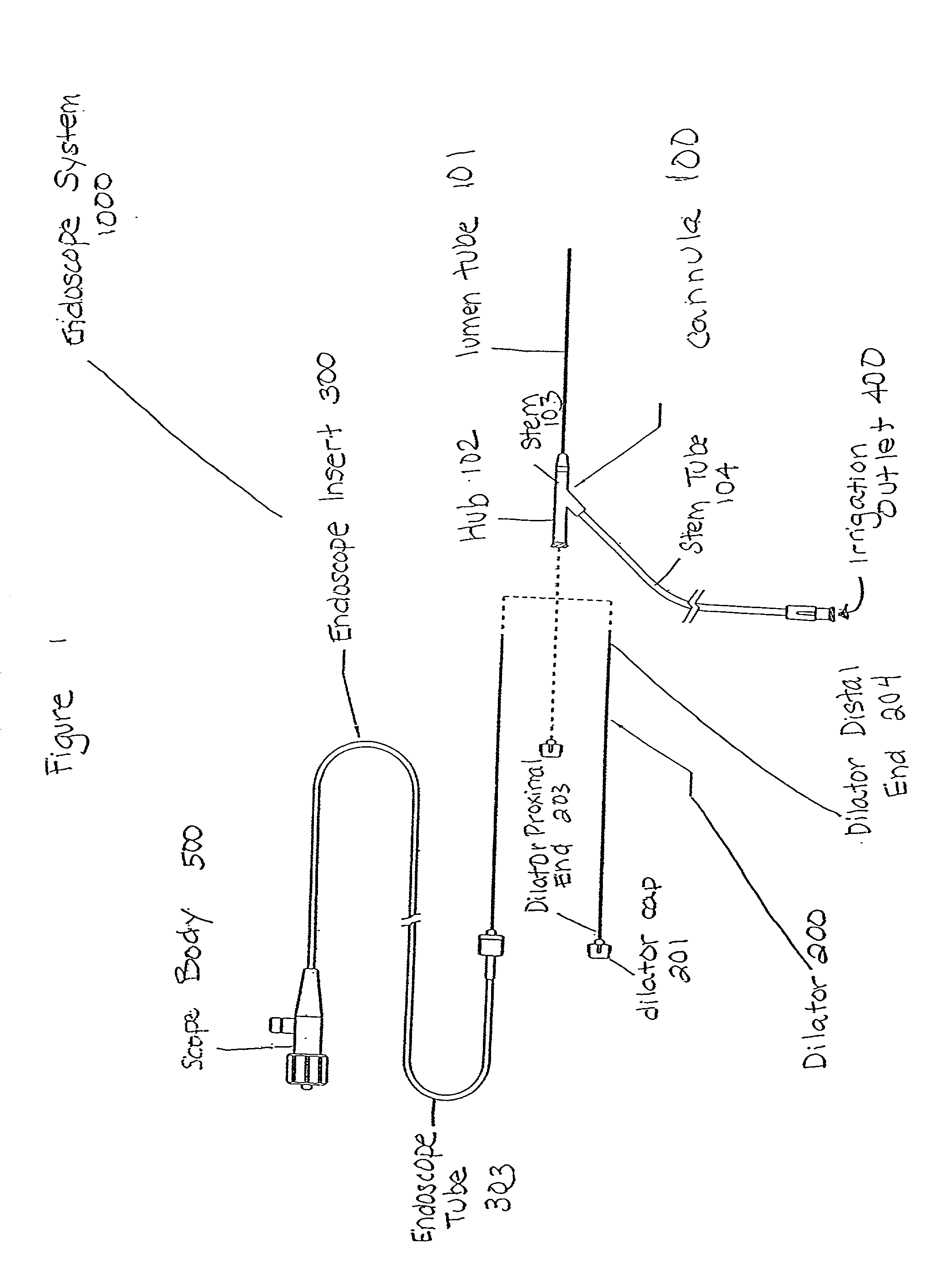
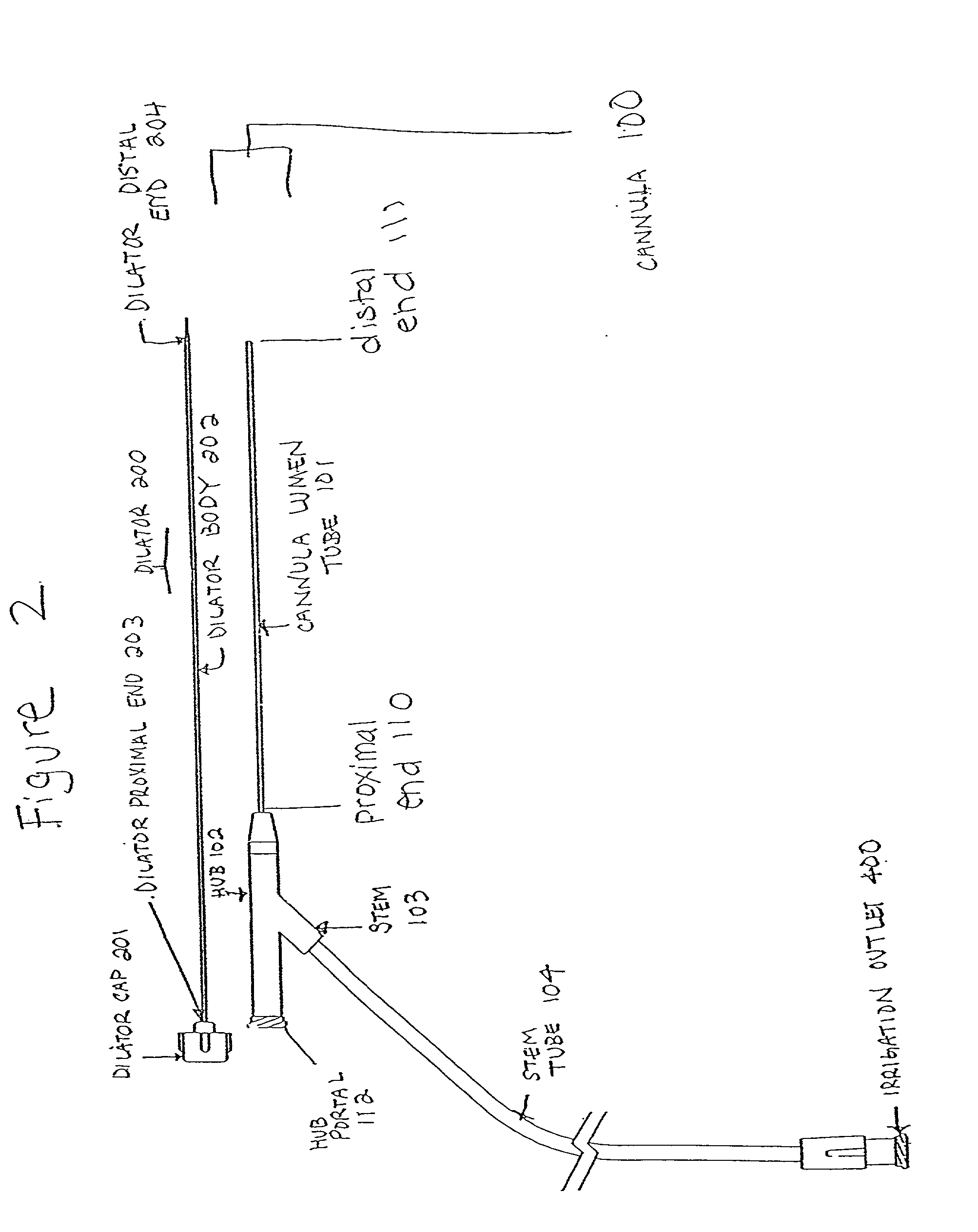
[0042] In endoscopic procedures, a video image is transmitted directly from the inside of the patient's body to a viewing device, such as a video monitor. Of particular importance is microendoscopic procedures that are viewed through micro-optic fiber image conduits. In breast exploration, microendoscopic procedures utilize scopes sized to be minimally invasive to breast tissues. These procedures utilize fiber optic bundles that range in diameter from 0.2 mm to 3.0 mm. Such procedures eliminate the need to open large operation areas and allow one to reach into and see inside very small and narrow body ducts. It reduces the patient's trauma, stress, danger of infection, allowing the patient in most cases to recover quickly.

[0043] Referring now to FIG. 1, the current endoscope system 1000 is comprised of...


PUM


Abstract

A three part microendoscope system including a cannula, a dilator, and endoscopic insert for detection of breast cancer are disclosed. To facilitate surgical entry of an endoscope system through the sphincter of a breast, a special dilator is used with an endoscope so that the endoscope can more readily pass through tortuous passageways. In accordance with the present invention, a semi-rigid endoscope can be advanced through the sphincter in the nipple using a nipple dilator. The microendoscope uses a special lumen tube that is usable with various insertable tools that allows for minimal invasive damage to surrounding cells. Insertable tools to irrigate, lavage, or perform biopsies on targeted areas of the breast are disclosed. A novel video coupler having a predetermined focus and orientation to be used with the present system is also disclosed. A method of using the three part microendoscope system and its various insertable tools are disclosed. A method of manufacturing the video coupler is disclosed.

Description

[0001] The present invention incorporates by reference in its entirety U.S. patent application Ser. No. 08 / 155,748, filed Nov. 23, 1993 by Tony Petitto and Stanislaw Loth, for "Technique for Depth of Field Viewing of Images With Improved Clarity and Contrast," now U.S. Pat. No. 5,400,177. It also incorporates by reference Ser. No. 08 / 115,748, filed Nov. 23, 1993, which is now U.S. Pat. No. 5,400,177, and Ser. No. 09 / 115,809, which is now abandoned, and Ser. No. 09 / 501,313, which is a continuation of Ser. No. 09 / 006,894, filed Jan. 14, 1998, and Ser. No. 09 / 524,746, filed Mar. 14, 2000.[0002] (1) Field of the Invention[0003] This invention relates generally to a medical apparatus and its medical use for diagnosing, exploring, and treating abnormalities in internal body tissue. In particular, the invention is directed to a three part endoscope system having a cannula, semi-rigid microendoscope, and a dilator. The present invention allows the use of a specifically designed microendosco...

Claims


Application Information

Patent Timeline
Patent Type & Authority Applications(United States)
IPC IPC(8): A61B1/00A61B1/04A61B1/07A61B1/12A61B18/24
CPCA61B1/04A61B1/07A61B1/12A61B18/24A61B1/00126A61B1/00167
Inventor GATTO, DOMINICK L.BONNEVILLE, JAMESJACENE, MICHAEL F.
Owner LACHOWICZ THEODORE COLLATERAL AGENT
Why Eureka
Try Eureka