Bendable cutting device

a cutting device and bendable technology, applied in the field of bendable cutting devices, can solve the problems of not being able to adapt to use in a minimally invasive procedure, not being able to directly visualize the actual procedure, and not being able to excise lesions of different sizes

Inactive Publication Date: 2004-11-04
ACUEITY HEALTHCARE
View PDF19 Cites 115 Cited by
  • Summary
  • Abstract
  • Description
  • Claims
  • Application Information

AI Technical Summary

Problems solved by technology

Devices that are typically used during open surgical procedures (i.e., scalpel, scissors, electrosurgical "pencil" electrode) are often not adaptable for use in a minimally invasive procedure.
In addition, the actual procedure cannot be directly visualized as the skin incision is typically just large enough to insert the device.
Current devices that use an oscillating sharp edge or radio frequency energy to cut the tissue retrieve a specimen of fixed volume and are not adaptable to excise lesions of different sizes or include extensions of the lesion or disease process in the excision.
Current minimally invasive devices are designed to excise a mass and are not adaptable for excision of an associated diseased duct or Cooper's ligament.
Leaving cancer behind in the duct or in Cooper's ligament increases the risk of local recurrence despite the administration of post operative radiation therapy.
Open surgical biopsy removes lesions of variable size and may include extensions of the lesion but often an excessive amount of normal breast tissue is included in the specimen leading to a poor cosmetic result.
In addition, open surgical biopsy typically requires a significant skin incision resulting in a longer, permanent scar.
More importantly, a diseased duct and / or disease in Cooper's ligament are not detectable either by direct vision or by palpation during an open surgical procedure.
The main cancerous mass may be excised, but a diseased duct filled with cancerous cells and / or diseased Cooper's ligament is often not appreciated during the procedure and unintentionally not included in the excision.

Method used


Image

  • Bendable cutting device
  • Bendable cutting device
  • Bendable cutting device

Examples


Embodiment Construction

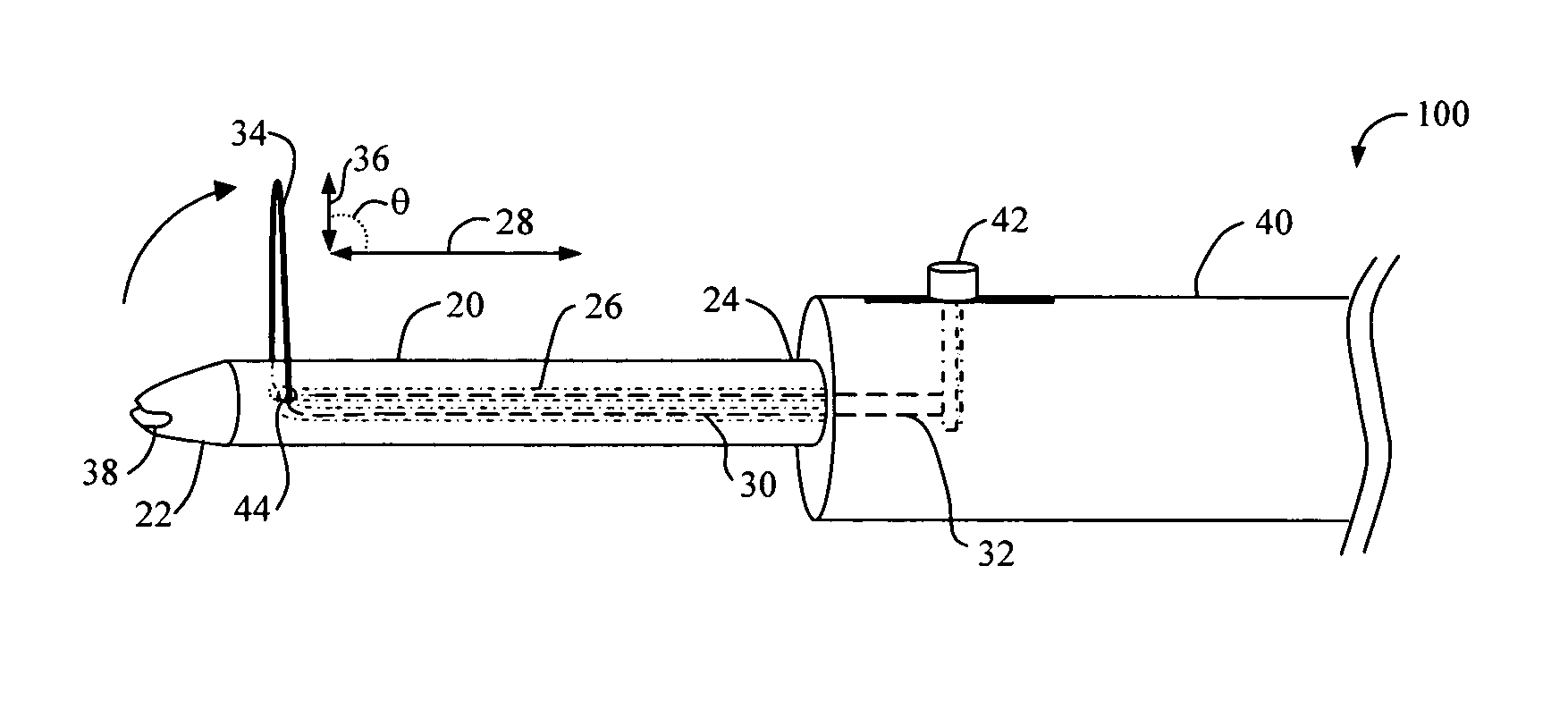
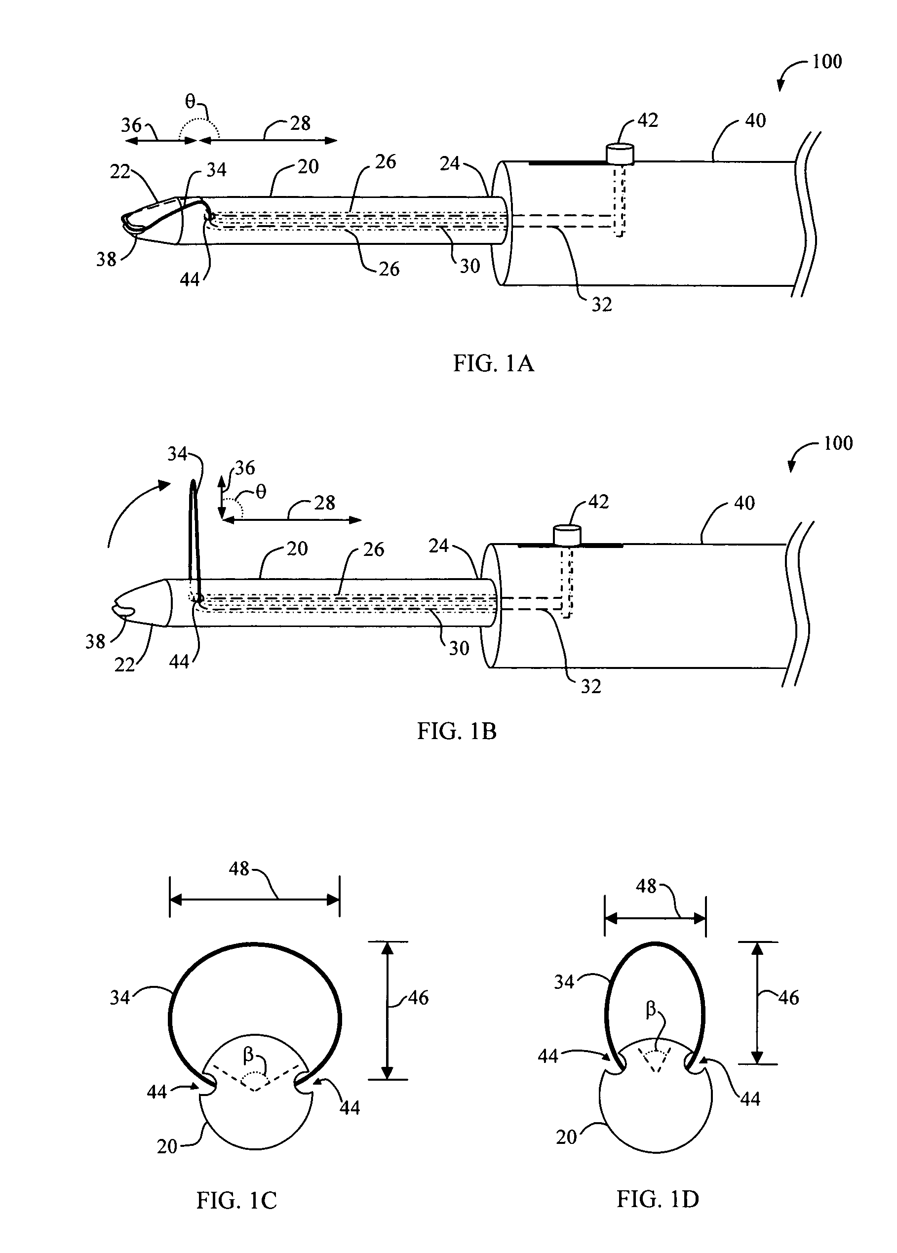
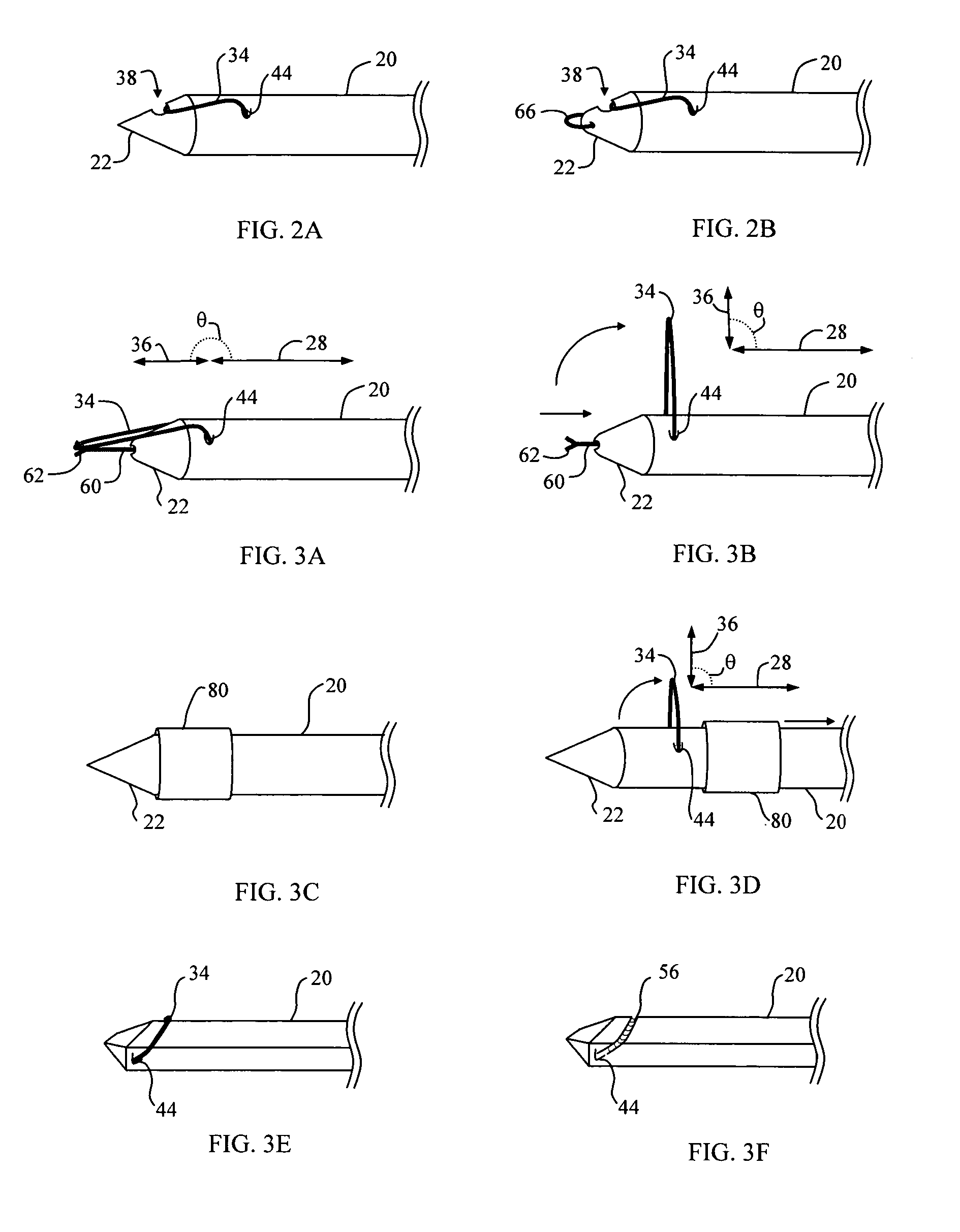
[0024] Devices and methods for a minimally invasive procedure for cutting or excising a volume of soft tissue such as a biopsy or a therapeutic excision of cancer are disclosed. The following description is presented to enable any person skilled in the art to make and use the invention. Descriptions of specific embodiments and applications are provided only as examples and various modifications will be readily apparent to those skilled in the art. The general principles defined herein may be applied to other embodiments and applications without departing from the spirit and scope of the invention. Thus, the present invention is to be accorded the widest scope encompassing numerous alternatives, modifications and equivalents consistent with the principles and features disclosed herein. For purpose of clarity, details relating to technical material that is known in the technical fields related to the invention have not been described in detail so as not to unnecessarily obscure the pr...


PUM


Abstract

Devices and methods for a minimally invasive procedure for cutting tissue are disclosed. The device generally includes a probe with a distal exit at an exit angle relative to the probe, a cutting loop with shape memory having a preconfigured shape, and a loop securing mechanism to secure the cutting loop in a penetrating configuration and to release the cutting loop into a cutting configuration. The cutting loop is generally within a profile of the probe in the penetrating configuration. In the cutting configuration, the cutting loop extends through the cutting loop exit and generally returns to the preconfigured shape at a cutting angle generally defined by the exit angle. The cutting loop securing mechanism may be, for example, a cover slidable over the probe, a slidable member extending from a distal tip of the probe, or a groove defined in the probe proximal to the cutting loop exit.

Description

[0001] This application claims priority to U.S. Provisional Patent Application Serial No. 60 / 449,061 filed on Feb. 20, 2003 which is incorporated herein by reference in its entirety.[0002] 1. Field of the Invention[0003] The present invention relates generally to devices and methods for cutting soft tissue. More specifically, devices and methods for a minimally invasive procedure for cutting or excising a volume of soft tissue such as a biopsy or a therapeutic excision of cancer are disclosed.[0004] 2. Description of Related Art[0005] Minimally invasive procedures have instigated a need for refinement in surgical devices that can function within confined spaces, particularly in soft tissue, such as breast tissue. Devices that are typically used during open surgical procedures (i.e., scalpel, scissors, electrosurgical "pencil" electrode) are often not adaptable for use in a minimally invasive procedure. In addition, the actual procedure cannot be directly visualized as the skin incis...

Claims


Application Information

Patent Timeline
Patent Type & Authority Applications(United States)
IPC IPC(8): A61B8/12A61B10/00A61B10/02A61B10/04A61B17/00A61B17/32A61B18/14
CPCA61B8/12A61B10/02A61B10/0266A61B10/04A61B17/32A61B17/320016A61B17/32056A61B2017/00867A61B2018/00601A61B2018/1407A61B2018/144
Inventor HO, HUDDEE JACOBLEE, ROBERTAZUCKSWERT, SAMUEL E.CONSTON, STANLEY R.YAMAMOTO, RONALDALDEN, DON
Owner ACUEITY HEALTHCARE
Why Eureka
Try Eureka