Method and apparatus for controlling the radiation dose in the generation of x-ray images for lithotripsy

a technology for lithotripsy and x-ray images, which is applied in the field of controlling the radiation dose in the generation of x-ray images for lithotripsy, can solve the problems of image contrast and thus calculus recognizability, and achieve the effects of reducing the brightness of the x-ray image, increasing the dose rate and the operating voltage of the x-ray tube, and improving the image contras

Inactive Publication Date: 2005-01-06
SIEMENS AG
View PDF42 Cites 5 Cited by
  • Summary
  • Abstract
  • Description
  • Claims
  • Application Information

AI Technical Summary

Benefits of technology

[0011] The invention is based on the realization that shadowed and diagnostically unusable image regions unavoidably exist in x-ray images from the focus region of a lithotripter. In an “out-line” arrangement this component is the partially imaged coupler bellows (cushion), (through which a portion of the x-ray radiation passes) of the shockwave generator that effects an attenuation of the x-ray radiation. In an “in-line” arrangement, this component is the surroundings of an aperture diaphragm used for the x-ray radiation in the coupler bellows, and the additional water flow line, which are traversed by the x-ray radiation. These items reduce the brightness of the x-ray image with respect to the total image field and cause the automatic dose control used in the prior art to both increase the dose rate and the operating voltage of the x-ray tube. In addition to an increase of the dose, due to the lesser primary absorption of higher-energy x-ray quanta, this additionally leads to a worsening of the image contrast and thus the recognizability of the calculus.
[0012] To control the x-ray system, the invention utilizes for exposure control only those parts of the total image field that are diagnostically usable and of paramount interest, and that preferably lie outside of a shadowed area and that includes the immediate surroundings of the focus of the lithotripter. As a result, a specific underexposure (measured on the average brightness of the total image field) occurs that is nevertheless sufficient to show the primary diagnostically interesting portion of the total image field in good image quality. As a result of this measure, the radiation exposure of the patient is decidedly reduced without leading to a worsening of the reproduction quality in these diagnostically interesting regions.

Problems solved by technology

In addition to an increase of the dose, due to the lesser primary absorption of higher-energy x-ray quanta, this additionally leads to a worsening of the image contrast and thus the recognizability of the calculus.

Method used


Image

  • Method and apparatus for controlling the radiation dose in the generation of x-ray images for lithotripsy
  • Method and apparatus for controlling the radiation dose in the generation of x-ray images for lithotripsy
  • Method and apparatus for controlling the radiation dose in the generation of x-ray images for lithotripsy

Examples


Embodiment Construction

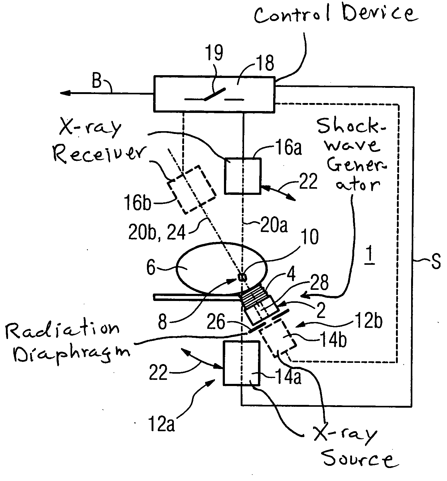
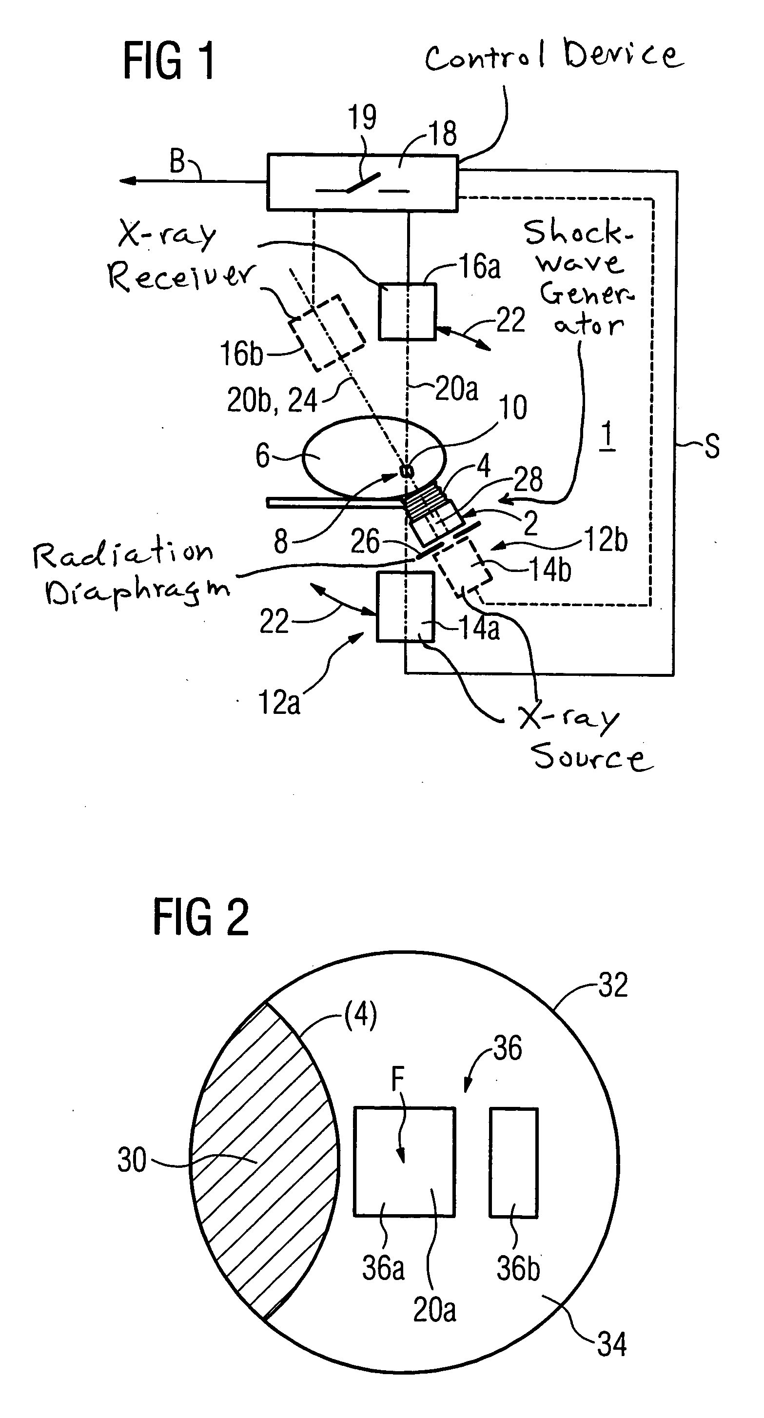
[0017] An apparatus according to the invention and shown schematically in FIG. 1 has a lithotripter 1 with a shockwave generator 2 placed over a coupler bellows 4 filled with water on the surface of the body 6 of a living subject. The shockwave generator 2 generates a shockwave that is focused in a focus region 8. Given correct positioning of the shockwave generator 2, a calculus 10 to be destroyed is located in this focus region 8.

[0018] An x-ray apparatus 12a that has an x-ray source 14a as well as an x-ray receiver 16a is associated with the lithotripter 1. A control signal S for inventive contrast-optimized and dose-optimized control of the x-ray source 14a is generated from the x-ray image acquired by the x-ray receiver 16a after digitization and evaluation. An optimized x-ray image B generated in this manner is output and, for example, reproduced on a monitor. With a selector switch 19 symbolically illustrated in FIG. 1, the user can decide whether a contrast-optimized and do...


PUM


Abstract

In a method and an apparatus to generate an x-ray image from the focus region of a lithotripter using x-rays that pass through the focus region and device parts of the lithotripter, to control the dose of the x-rays, only an image region of the image field exposed by the x-rays, that lies outside of an area shadowed by these device parts, is evaluated.

Description

BACKGROUND OF THE INVENTION [0001] 1. Field of the Invention [0002] The present invention concerns a method for controlling the radiation dose in the generation of an x-ray image from the focus region of a lithotripter, and an apparatus to implement the method. [0003] 2. Description of the Prior Art [0004] Lithotripsy is a therapeutic method to destroy (without a surgical procedure) a calculus located in the body of an organism using a focused ultrasound shockwave that is generated by a shockwave generator. In order to largely prevent damages in the tissue surrounding the calculus, it is necessary on the one hand to position the focus of the lithotripter exactly in the calculus. It is also necessary to visualize the progress in the destruction of the calculus in order to limit the number of shockwave pulses to the therapeutically necessary minimum. [0005] To generate an x-ray image of the environment of the focus region of a lithotripter, it is known, for example, from German OS 197...

Claims


Application Information

Patent Timeline
Patent Type & Authority Applications(United States)
IPC IPC(8): A61B6/00A61B17/22A61B19/00H05G1/26
CPCA61B17/2256A61B6/4441
Inventor KALTSCHMIDT, RAINER
Owner SIEMENS AG
Why Eureka
Try Eureka