

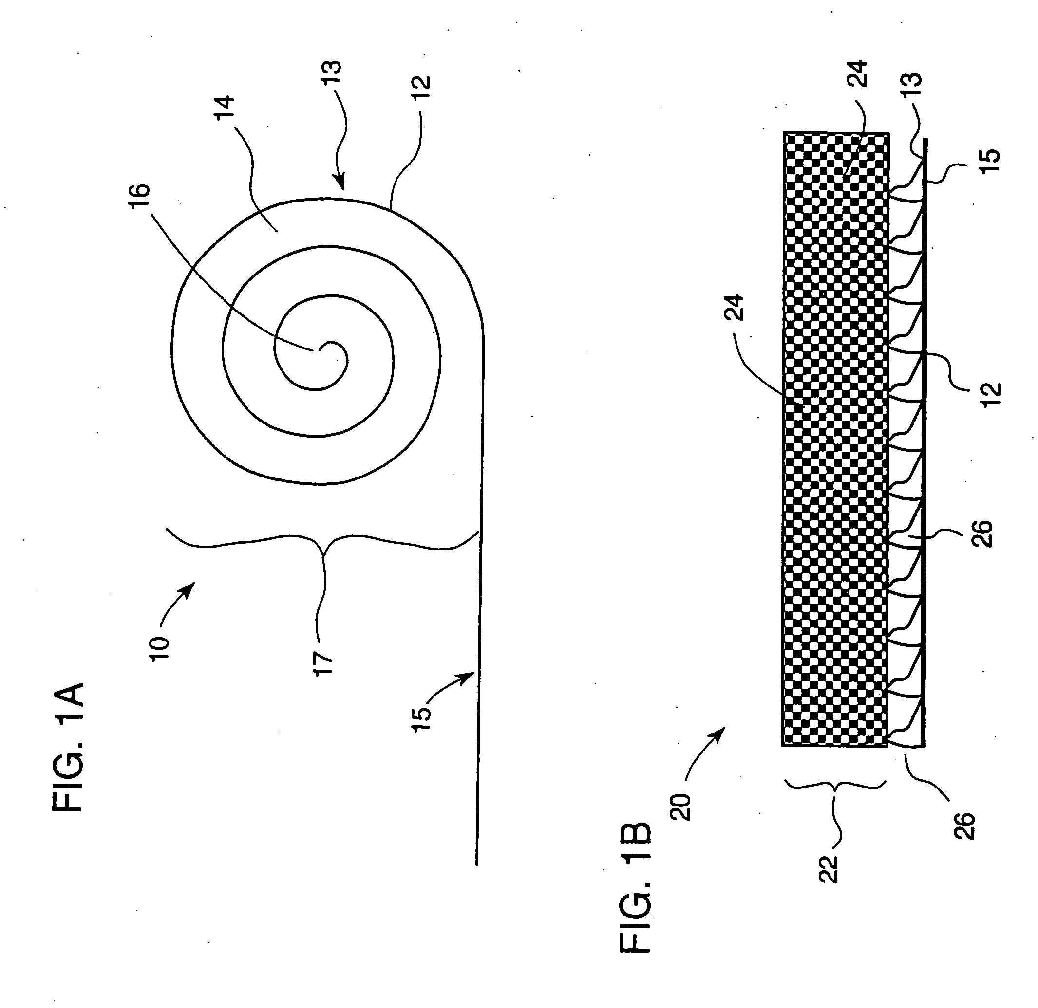
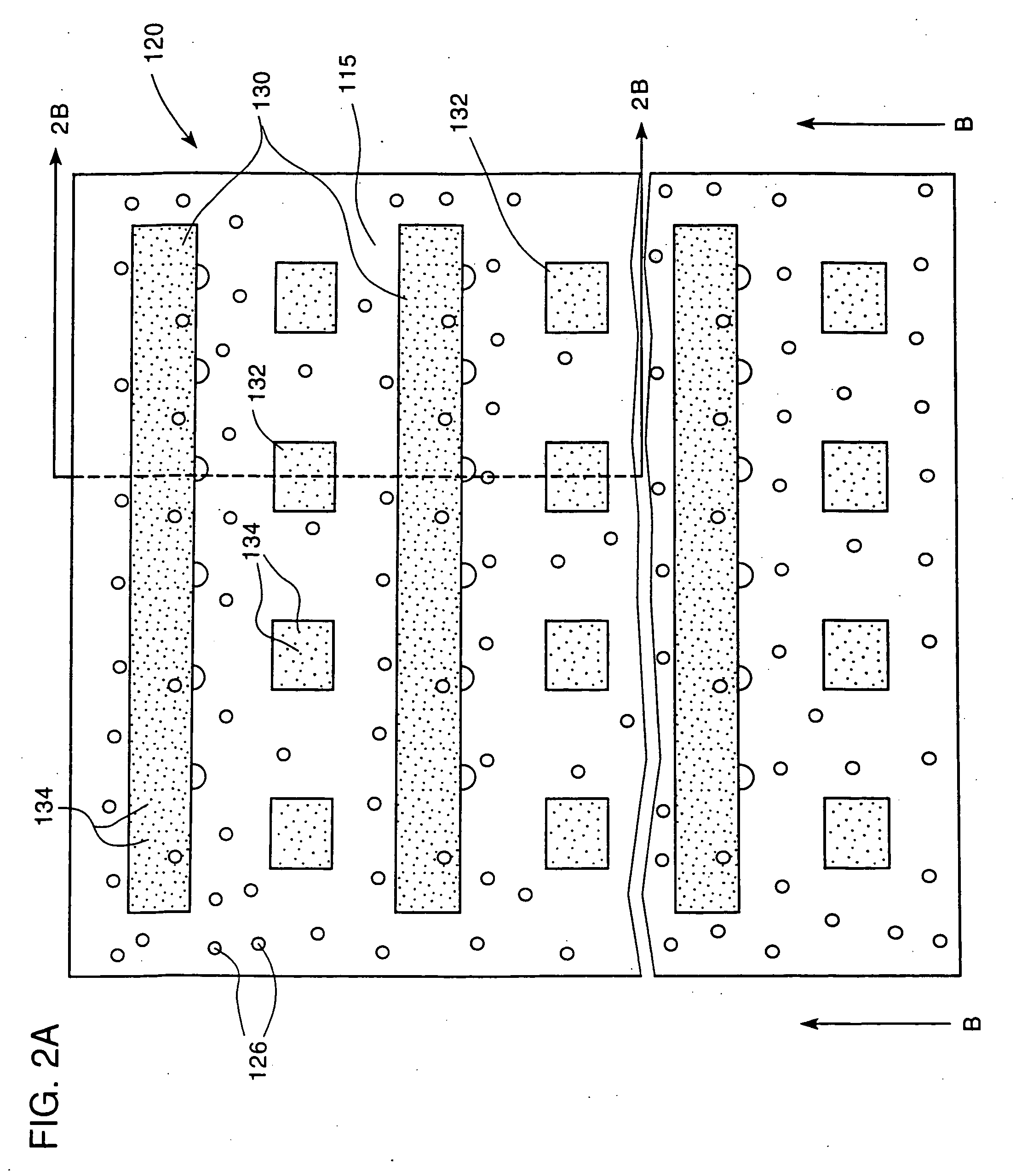
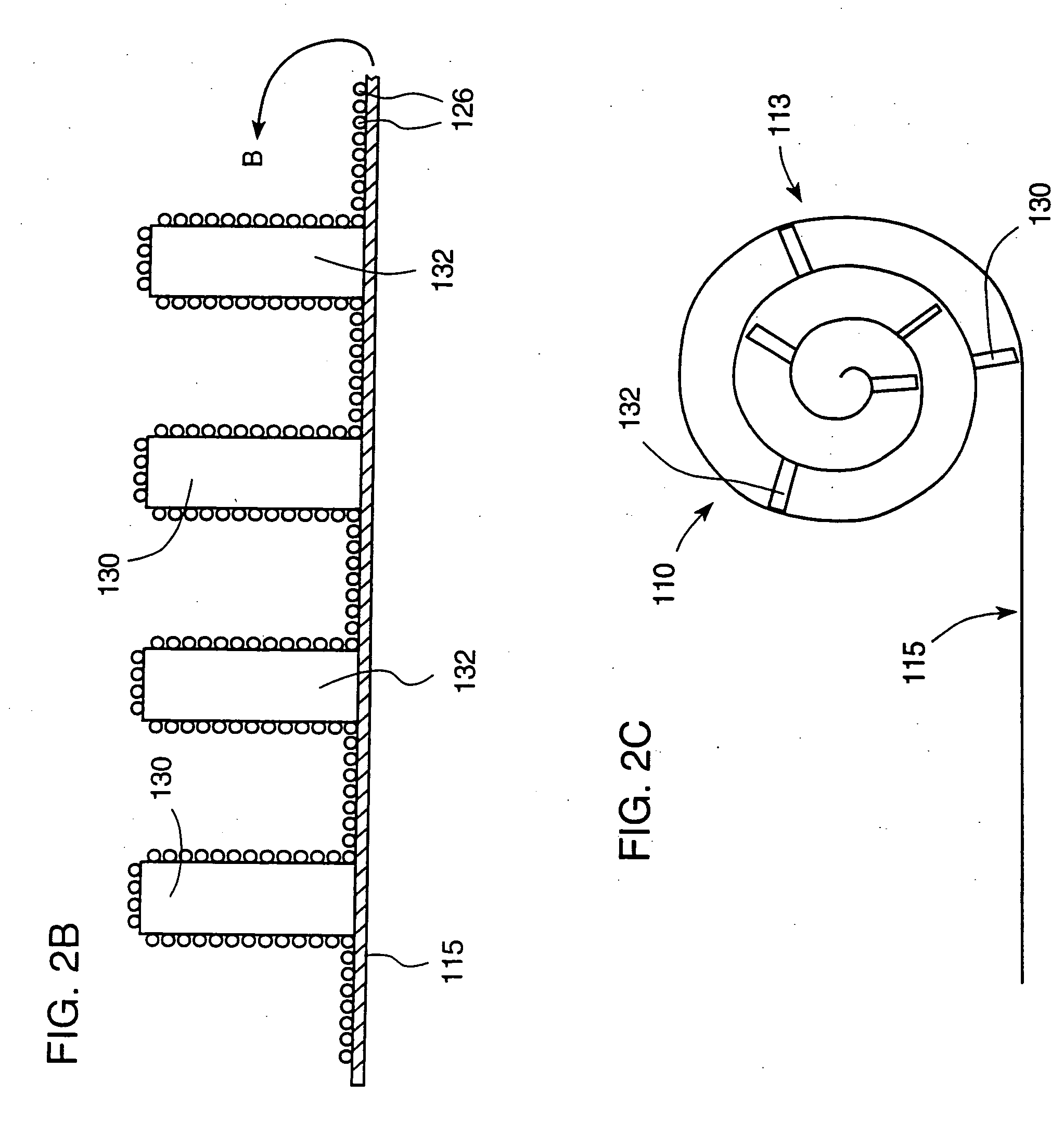
Neural regeneration conduit
a neurotrophic agent and conduit technology, applied in the field of can solve the problems of limited success in the combination of these approaches, and achieve the effect of facilitating neurotrophic agent concentration gradient formation
- Summary
- Abstract
- Description
- Claims
- Application Information
AI Technical Summary
Benefits of technology
Problems solved by technology
Method used
Image

Examples
example
[0046] Schwann cells were isolated from neonatal Fisher rats. Small intestinal submucosa (SIS) was harvested from adult Fisher rats for use as a support material in a nerve regeneration conduit. The SIS was cut into 7 mm by 8 cm pieces and pinned out. Schwann cells were plated onto the SIS sheets and cultured until they reached confluence. The strips were then rolled into a laminar structure and implanted across a 7 mm gap in the rat sciatic nerve (n=12). Control animals received SIS conduits without Schwann cells (n=11) or an autograft repair (n=12).
[0047] At both 6 and 10½ weeks, functional recovery through the Schwann cell-laden SIS conduits, measured by sciatic function index, exceeded that through the cell-free conduits, but compared favorably with autografts.
PUM
 Abstract
Description
Claims
Application Information