Neural cell assay
a technology of neuronal cells and assays, applied in the field of neurology, neurology, cell biology and toxicology, can solve the problems of brain function impairment, severe consequences, and impaired cognition and memory
- Summary
- Abstract
- Description
- Claims
- Application Information
AI Technical Summary
Benefits of technology
Problems solved by technology
Method used
Image
Examples
example 1
Isolating Brain Marrow from an Animal
[0071] A portion of an animal brain (e.g., RMS, SEZ, hippocampus) can be surgically removed and mechanically separated into smaller tissue pieces. One such method for mechanically separating a brain sample into smaller tissue pieces involves mincing the brain sample with a razor blade. To further dissociate the pieces into a single cell suspension, the tissue pieces are incubated in a solution containing a proteolytic enzyme such as trypsin, papain and / or hyaluronidase. An example of such an incubation involves incubating tissue pieces in a trypsin / EDTA solution for 10 minutes at 37° C. Other examples include an incubation in 14 U / ml of papain or 1.3 mg / ml trypsin and 0.67 mg / ml hyaluronidase for 1 hour with gentle rocking.
[0072] Following incubation with an enzyme-containing solution, the tissue may be subjected to further mechanical dissociation using a Pasteur pipette (e.g., fire-polished Pasteur pipette). Once the tissue is dissociated into...
example 2
Neurosphere Formation
[0073] Typically, isolated neural cells are cultured in a medium that permits the growth and proliferation of neurospheres. The culture in which the isolated cells proliferate can be a serum-free medium containing one or more predetermined growth factors effective for inducing multipotent neural stem cell proliferation. The culture medium can be supplemented with a growth factor selected from leukocyte inhibitory factor (LIF), epidermal growth factor (EGF), basic fibroblast growth factor (FGF-2; bFGF) or combinations thereof. The culture medium can be further supplemented with neural survival factor (NSF) (San Diego, Calif.) and / or fetal bovine serum. Neurospheres cultured according to this method are not immunoreactive for glial fibrillary acidic protein (GFAP; a marker for astrocytes), neurofilament (NF; a marker for neurons), neuron-specific enolase (NSE; a marker for neurons) or myelin basic protein (MBP; a marker for oligodendrocytes). However, cells withi...
example 3
Analyzing Effects Of Agents on Brain Cells
[0075] Certain brain marrow constituents have been previously characterized. It has been established that the periventricular subependymal zone (SEZ) of the adult mouse brain exhibits persistent expression of developmentally regulated molecules, including the extracellular matrix (ECM) protein tenascin that delineates the rostral migratory stream (RMS), extending from the lateral ventricle to the olfactory bulb (OB). This dense ECM within the brain marrow, similar to what is observed in bone marrow, can be visualized in brain sections, for example using an antibody to tenascin. Marrow tissues are characterized as exhibiting persistent cell proliferation, which in the case of brain marrow reflects ongoing neurogenesis.
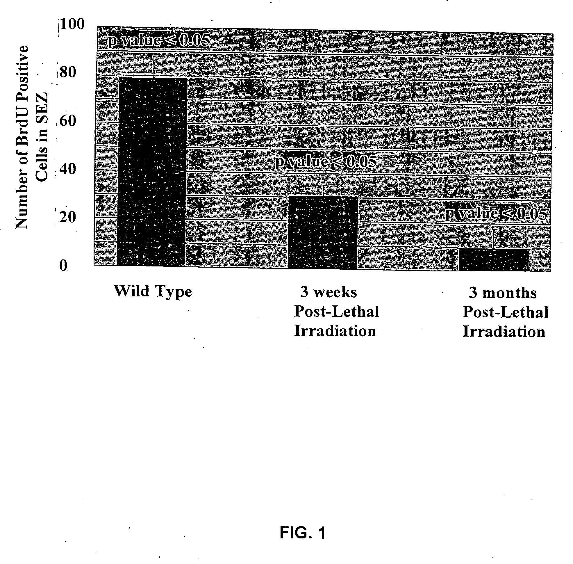
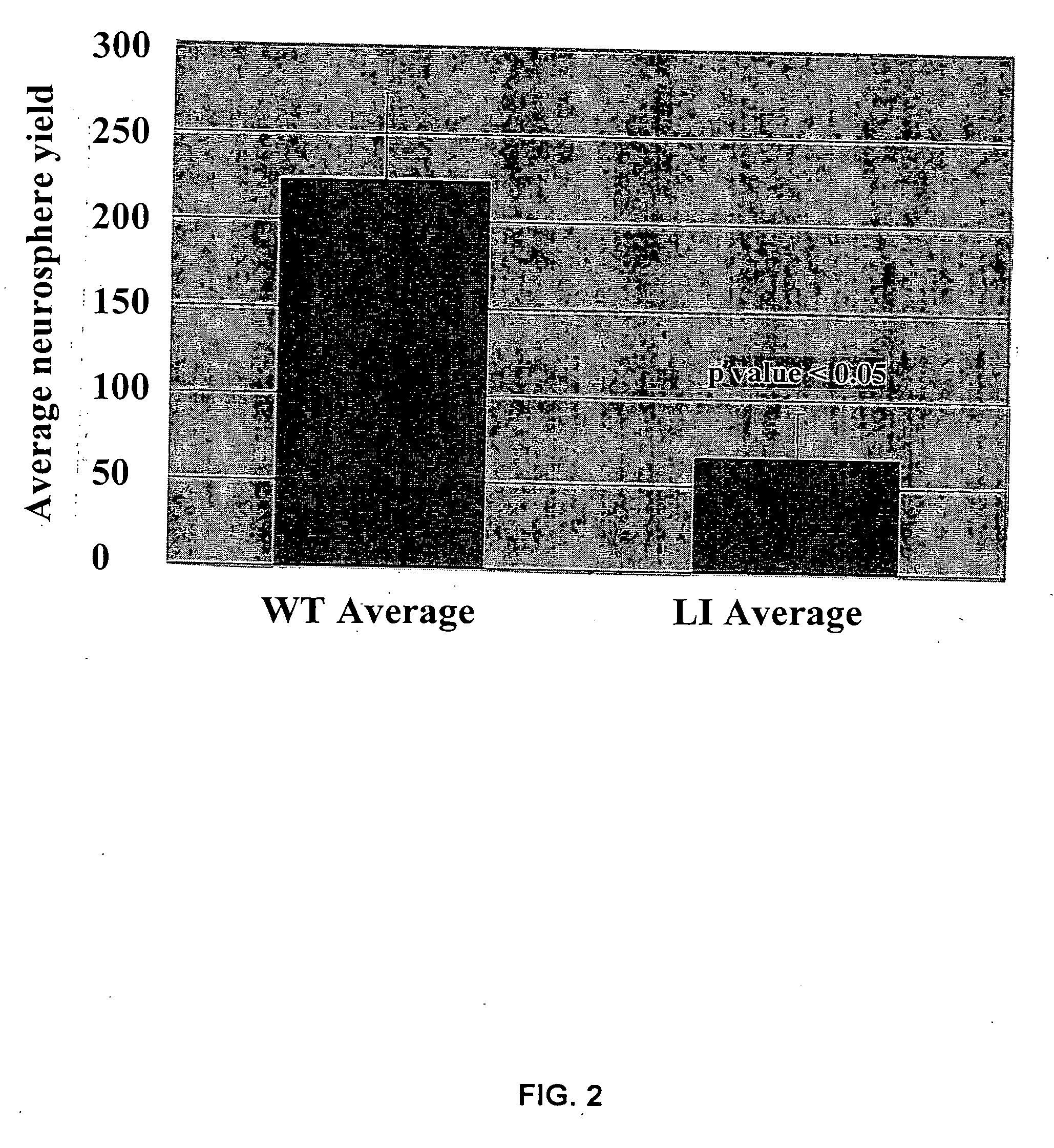
[0076] Immunostaining of sagittal brain sections including the SEZ from control adult mice using an antibody against neuronal β-III tubulin revealed densely packed immunopositive immature neurons having few processes, oriented...
PUM
| Property | Measurement | Unit |
|---|---|---|
| volume | aaaaa | aaaaa |
| volume | aaaaa | aaaaa |
| volume | aaaaa | aaaaa |
Abstract
Description
Claims
Application Information
 - R&D
- Intellectual Property
- Life Sciences
- Materials
- Tech Scout
- Unparalleled Data Quality
- Higher Quality Content
- 60% Fewer Hallucinations
Browse by: Latest US Patents, China's latest patents, Technical Efficacy Thesaurus, Application Domain, Technology Topic, Popular Technical Reports.
© 2025 PatSnap. All rights reserved.Legal|Privacy policy|Modern Slavery Act Transparency Statement|Sitemap|About US| Contact US: help@patsnap.com


