Products and methods for single parameter and multiparameter phenotyping of cells

Inactive Publication Date: 2005-04-28
HEMATOTYPES
View PDF38 Cites 1 Cited by
  • Summary
  • Abstract
  • Description
  • Claims
  • Application Information

AI Technical Summary

Benefits of technology

[0012] A method of characterizing cells is provided, comprising the steps of a) providing a suspension of cells in a liquid medium, said cells including first cells, b) adding to said suspension a group of substantially identical first beads, each of said first beads being coated with a binding substance or being magnetic such that each first bead is adapted to bind to a first cell, c) incubating said first beads in said suspension for a period of time effective to permit said first beads to bind to said first cells to form first bead-first cell complexes, each first bead-first cell complex comprising a first bead and a first cell, d) separating said first bead-first cell complexes from said suspension by filtration, and e) examining

Problems solved by technology

However, currently available methodologies, particularly flow cytometry, are expensive and require a high degree of suspicion at the time of biopsy.
If tissue is not set aside and there is cancer present, the correct subtyping of the tumor (and proper assignment to treatment protocols) cannot be done after the fact.
Methods that do not require forethought, such as immunostaining of paraffin blocks, are far less sensitive and do not work well in laboratories that do not perform these stains frequently.
However, there are several problems with the method.
The expense of establishing and maintaining these laboratories is perhaps the most severe problem.
Uncontrollable factors such as temperature variations, rough handling, bacterial contamination, or shipping delays may render samples unsuitable for analysis.
In addition, flow cytometry requires technologists who have specialized training and their time must often be dedicated solely to the technology itself, further increasing the expense of the procedure.
This is because the number of red cells in blood and bone marrow samples is far greater than other cells types, and shear numbers alone would overwhelm the sensitive detectors of the machines.
This method virtually eliminates megakaryocytes from most analysis and frequently destroys delicate malignant cells (particularly from the relatively common tumors such as large cell lymphoma and Hodgkin's disease).
It is in these situations that the great sensitivity and complexity of flow cytometry may work to its disadvantage.
Without the information that flow cy

Method used


Image

  • Products and methods for single parameter and multiparameter phenotyping of cells
  • Products and methods for single parameter and multiparameter phenotyping of cells

Examples


example 1

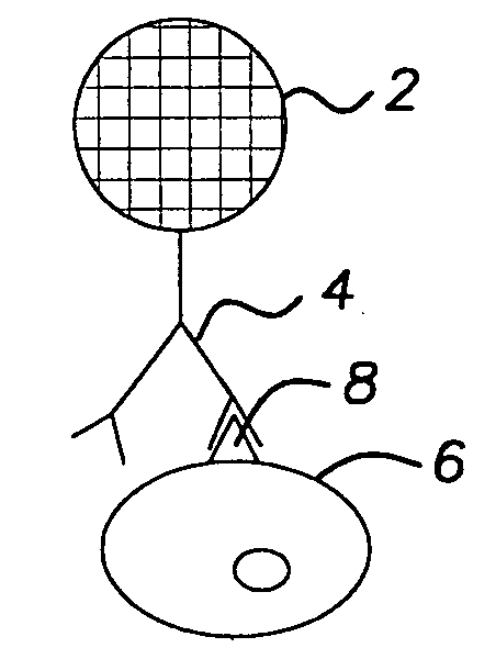
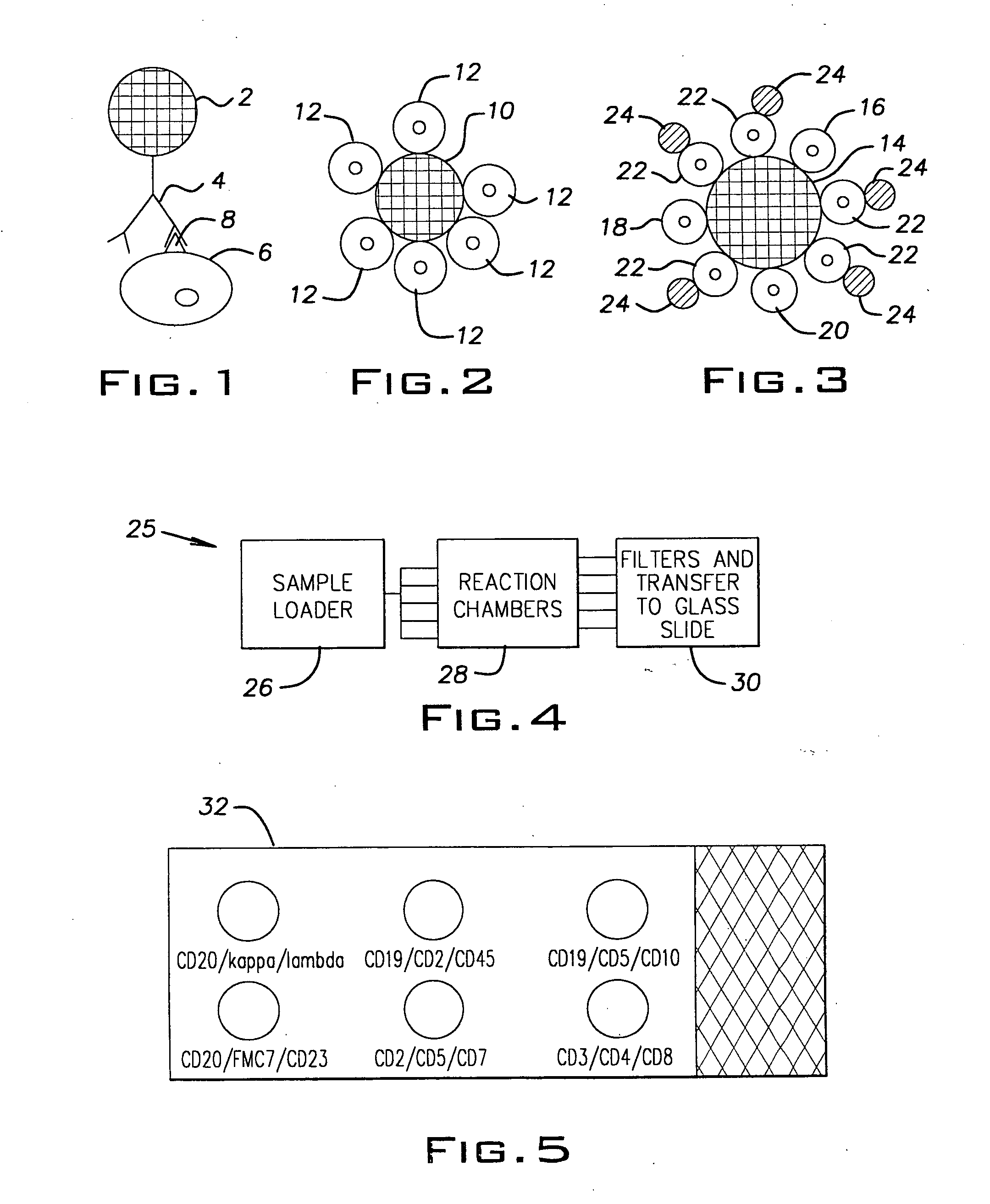
[0049] A 30 year old man presented with pancytopenia and splenomegaly. Examination of the peripheral smear confirmed the pancytopenia. In addition, scattered cells were present that showed bland cytological characteristics, with a monocytoid appearance. The nuclei of these cells were round to oval, with a single intermediate nucleolus. There was abundant blue-gray cytoplasm that showed numerous cytoplasmic projections. A bone marrow examination revealed a hypocellular aspirate with similar cells present. Small clusters of abnormal cells were present on the core biopsy. A buffy coat sample of the peripheral smear was suspended in anti-CD20 coated 10-micron colorless beads to distinguish the abnormal cells from monocytes. The suspension was passed through an appropriate filter and the cells were then transferred to a glass slide and stained. A schematic of the resulting slide preparation is demonstrated in FIG. 2. Positive binding of the abnormal cell population to the 10-micron beads...

example 2

[0050] A 68 year old man with a known history of chronic lymphocytic leukemia (CLL) presented for routine follow up examination. Clinical examination revealed that the patient had a peripheral white cell count of 435,500 cells / ml (normal range 4,300-11,000 cells / ml) which included 87% lymphocytes. Morphologic examination of the peripheral blood smear revealed predominantly an abnormal population of small lymphocytes with a small but significant population of large transformed cells. A suspension of cells in a liquid medium was provided. This sample was analyzed using anti-CD20 coated 10-micron beads, anti-kappa coated colorless 5-micron beads and anti-lambda coated colorless 5-micron beads in two separate tubes. In the procedure, the same sample was placed into each of 2 tubes. To each tube was added anti-CD20 coated 10-micron beads. These strongly bound the B cells. The question then was whether the B cells were kappa, lambda or a combination of both. Therefore, the 5 micron anti-k...

example 3

[0051] A 19 year old man presented with headache and stiff neck to the emergency. His evaluation included obtaining a sample of cerebral spinal fluid for which emergency pathologist evaluation of the fluid was requested to rule out the presence of “blasts”. Evaluation showed a relatively uniform population of small lymphocytes, and a diagnosis of viral meningitis was suggested. The patient's physician requested flow cytometry to completely rule out the possibility of malignancy. Since excess fluid was available, a small sample was treated with anti-CD20 coated 10-micron beads and anti-kappa and anti-lambda coated 5-micron beads in two separate tubes using essentially the same procedure as described in Example 2 above. The majority of cells did not bind to either the anti-CD20, anti-kappa, or anti-lambda beads, suggesting that the lymphoid population was composed predominantly of T cells. Flow cytometric analysis received two days later confirmed approximately 60% T cells and 40% B c...


PUM

PropertyMeasurementUnit
Pore sizeaaaaaaaaaa
Pore sizeaaaaaaaaaa
Diameteraaaaaaaaaa

Abstract

A method of single parameter and multiparameter characterizing of cells, particularly immunophenotyping of cells, is provided. The method preferably uses antibody coated microspheres which are adapted to bind to specific types of cells. One or more sets of coated microspheres are added simultaneously or sequentially to a suspension of cells and bind the cells they are adapted to bind. Cells may bind to one or more microspheres. The suspension is then filtered to trap bead-cell complexes. The complexes are preferably stained and then examined to characterize the cells, preferably the cells bound to the microspheres. A kit and apparatus for performing the method are also provided.

Description

[0001] This application claims the benefit of U.S. Provisional Application Ser. No. 60 / 132,395 filed May 4, 1999. The contents of said Provisional Application Ser. No. 60 / 132,395 are incorporated herein by reference in their entirety.FIELD OF THE INVENTION [0002] The present invention relates generally to phenotyping and immunophenotyping of cells and more particularly to single parameter and multiparameter phenotyping and immunophenotyping of cells. BACKGROUND OF THE INVENTION [0003] Immunophenotyping of cells and tumors, particularly hematopoietic tumors, is often of critical importance for clinical evaluation of cancer patients. However, currently available methodologies, particularly flow cytometry, are expensive and require a high degree of suspicion at the time of biopsy. All too often, even before the diagnosis of cancer is made, precious tissue must be set aside for possible immunophenotyping. If tissue is not set aside and there is cancer present, the correct subtyping of t...

Claims


Application Information

Patent Timeline
IPC IPC(8): G01N33/569
CPCG01N33/54313G01N33/56966G01N33/57426Y10S436/824Y10S435/961Y10T436/255Y10T436/2525Y10T436/25375Y10T436/101666Y10T436/25125Y10T436/25
Inventor PANKOWSKY, DAN A.
Owner HEMATOTYPES
Why Eureka
Try Eureka