
Determination of disease state using Raman Spectroscopy of tissue

Inactive Publication Date: 2005-04-28
VERALIGHT INC
View PDF41 Cites 32 Cited by
  • Summary
  • Abstract
  • Description
  • Claims
  • Application Information

AI Technical Summary

Benefits of technology

[0019]“Tissue reflectance characteristic” includes any reflectance property of tissue that is useful in correction of detected light, including as examples the tissue reflectance at the Raman excitation wavelength, the tissue reflectance at the Raman scattering wavelength, and the tissue reflectance at other wavelengths found useful for estimating the tissue's intrinsic Raman scattering spectrum. A “measure of chemical change due to glycemic control” means any change in the chemical characteristics of tissue that is due to glycemic control, examples including concentration, measurements of the presence, concentration, or change in concentration of glycation end-products in tissue; measurements of the rate or change in the rate of the accumulation of such end-products; measurements of tissue membrane thickness or the change, rate of change, or direction of change of such thickness; tissue properties such as tensile strength, strain, or compressibility, or the change, rate of change, or direction of change of such property. A “measure of glycation end-product” means any measure of the presence, time, extent, or state of tissue associated with hyperglycemia, including, as examples, measurements of the presence, concentration, or change in concentration of glycation end-products in tissue; measurements of the rate or change in the rate of the accumulation of such end-products; measurements of the presence, intensity, or change in intensity of Raman scattering signal at Stokes shifts known to be associated with tissue glycation end-products; and measurements of the rate or change in the rate of the accumulation of such Raman signal. When light is described as having a “single wavelength,” it is understood that the light can actually comprise light at a plurality of wavelengths, but that a significant portion of the energy in the light is transmitted at a single wavelength or at a range of wavelengths near a single wavelength.

Problems solved by technology

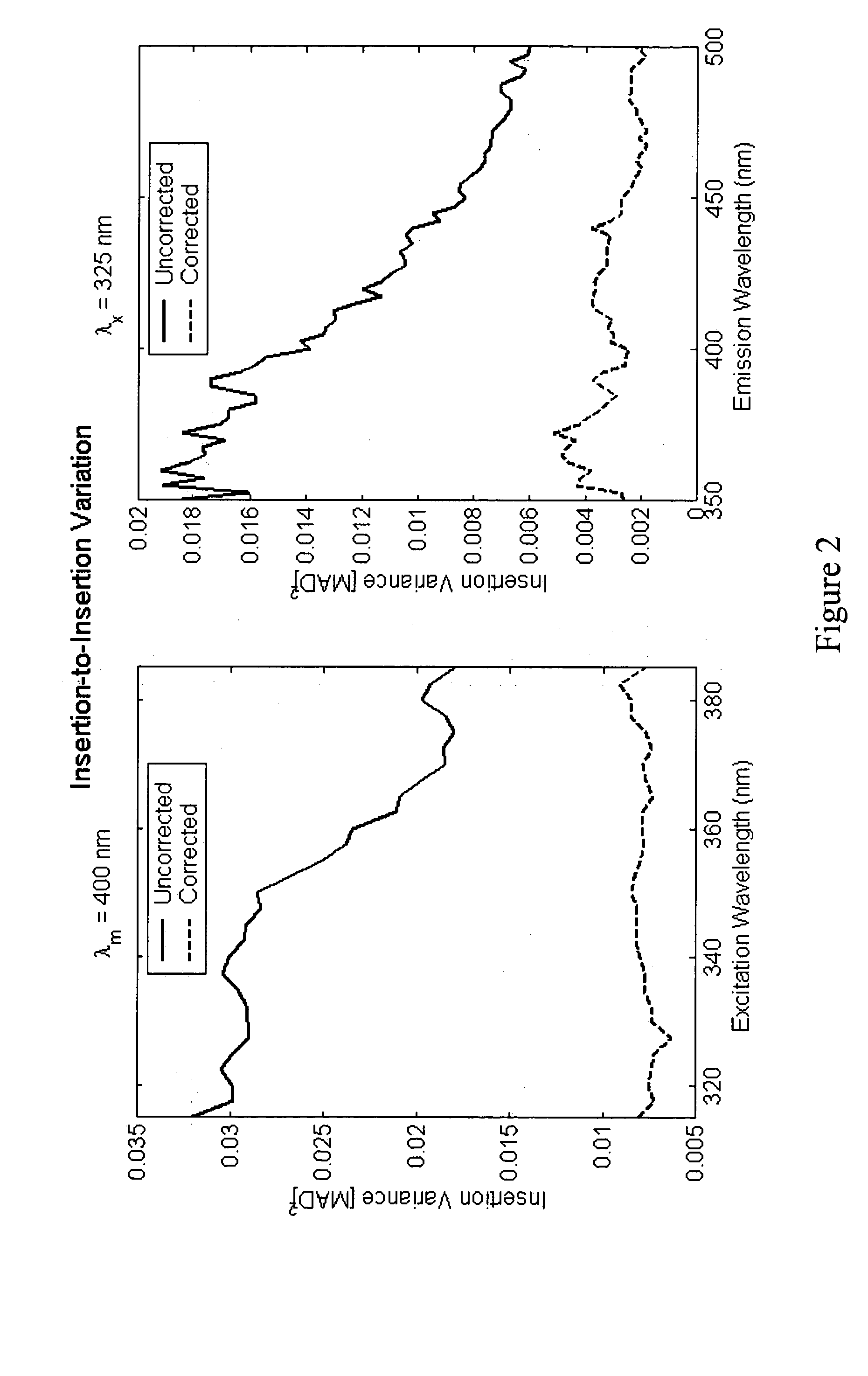
For example, the reflectance of the tissue can lead to errors if appropriate correction is not employed.

Method used


Image

  • Determination of disease state using Raman Spectroscopy of tissue
  • Determination of disease state using Raman Spectroscopy of tissue
  • Determination of disease state using Raman Spectroscopy of tissue

Examples


example i

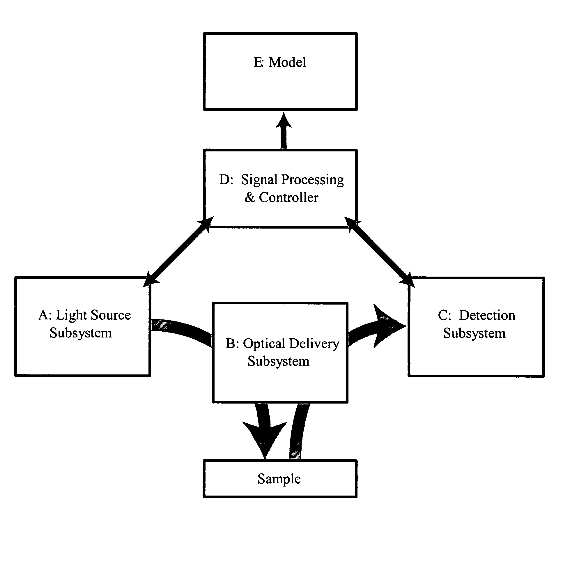
[0057] Example I of such a system embodies a continuous-wave diode laser as the light source. The optical-coupling sub-system is comprised of a fiber probe that couples the excitation light to the tissue and collects Raman scattering emanating from the tissue, as illustrated in FIG. 6. The probe incorporates a line-pass filter to reject Raman emission of the fiber itself, as well as rejecting any broad spontaneous emission from the diode laser (i.e., the filter only passes the narrow bandwidth of the laser). A pair of dichroic mirrors creates a coaxial path for excitation and emission. A final lens focuses the excitation radiation and then collects and collimates Raman emission. The Raman emission passes through the second of the dichroic pair and a line blocking filter rejects Rayleigh scattered excitation final. A lens couples the Raman emission into a fiber or fiber bundle for relay to the detection system. The detection system contains a spectrograph and a detector such as a CCD...


PUM


Abstract

A method of determining disease state in an individual. A portion of the tissue of the individual is illuminated with excitation light, then light emitted by the tissue due to Raman scattering of a chemical with the tissue responsive to the excitation light is detected. The detected light can be combined with a model relating Raman emission with disease state to determine a disease state of the individual. The invention can comprise single wavelength excitation light, scanning of excitation light (illuminating the tissue at a plurality of wavelengths), detection at a single wavelength, scanning of detection wavelengths (detecting emitted light at a plurality of wavelengths), and combinations thereof. The invention also can comprise correction techniques that reduce determination errors due to detection of light other than that from Raman emission of a chemical in the tissue. For example, the reflectance of the tissue can lead to errors if appropriate correction is not employed. The invention can also comprise a variety of models relating Raman emission to disease state, including a variety of methods for generating such models. Other biologic information can be used in combination with the Raman spectral properties to aid in the determination of disease state. The invention also comprises apparatuses suitable for carrying out the method, including appropriate light sources, detectors, and models (for example, implemented on computers) used to relate detected Raman emission and disease state.

Description

CROSS REFERENCES TO CO-PENDING APPLICATIONS [0001] This application claims priority under 35 U.S.C § 120 to U.S. patent application Ser. No. 10 / 116,272, entitled “Apparatus And Method For Spectroscopic Analysis Of Tissue To Detect Diabetes In An Individual,” filed Apr. 4, 2002, incorporated herein by reference. This application claims priority under 35 U.S.C § 119 to U.S. provisional application 60 / 515,343, Determination of a Measure of a Glycation End-Product or Disease State Using Tissue Fluorescence,” filed Oct. 28, 2003, and U.S. provisional application 60 / 517,418, Apparatus and Method for Spectroscopic Analysis of Tissue to Determine Glycation End-products incorporated herein by reference.FIELD OF THE INVENTION [0002] The present invention generally relates to determination of disease state from Raman spectroscopy in tissue. More specifically, the present invention relates to methods and apparatuses for determining models that relate Raman spectra of tissue to disease state, an...

Claims


Application Information

Patent Timeline
Patent Type & Authority Applications(United States)
IPC IPC(8): A61B5/00A61B5/103
CPCA61B5/0059A61B5/0064A61B5/0066A61B5/0068A61B5/443A61B5/0075A61B5/061A61B5/412A61B5/441A61B5/0071
Inventor EDIGER, MARWOOD NEALGARDNER, CRAIG M.HULL, EDWARD L.
Owner VERALIGHT INC
Learn More