Left atrial access apparatus and methods

a technology of access apparatus and left atrial cavity, which is applied in the field of medical devices, can solve the problems of serious injury or death of patients, complication of transeptal puncture, and inability to fully fully cover the aorta or the free wall of the atrium

Inactive Publication Date: 2005-05-26
HANSEN MEDICAL INC
View PDF20 Cites 26 Cited by
  • Summary
  • Abstract
  • Description
  • Claims
  • Application Information

AI Technical Summary

Benefits of technology

[0009] To gain access to the left atrium within the heart, a transeptal puncture may be performed by a clinician utilizing a sounding procedure. Such a procedure may take advantage of physiological parameter differences between the left and right atrial chambers to detect a direct pathway therebetween. For example, one method may be to utilize the oxygen saturation difference between the atrial chambers in guiding a needle or catheter into the appropriate position.

Problems solved by technology

If the targeted area is missed by the penetrating device, structures such as the aorta or the free wall of the atrium may be in danger of being penetrated.
If such a penetration were to occur, serious injury or death to the patient could result.
In fact, the most common serious complication of transeptal puncture is cardiac tamponade, a life-threatening condition resulting from misplacement of the transeptal puncture leading to bleeding into the pericardial space.
However, fluoroscopy provides only two-dimensional imaging and does not preclude the possibility of the clinician inadvertently puncturing the septum in the wrong location.
Some reports have shown that trans-esophageal echocardiography and intracardiac echocardiography can improve the safety of transeptal puncture; however these additional procedures are expensive, inconvenient, and add further risk to the procedure.
Perfecting the technique of transeptal puncture is very challenging for the clinician.
Very few interventional cardiologists and electrophysiologists are currently skilled at this procedure.
As difficult as it is to perform, it is more difficult to teach and relies upon both visual and tactile senses.
Obtaining safe access to the left atrium remains a major obstacle to the growth of the technologies relating to the left atrium, e.g., left-sided arrhythmia mapping and ablation, atrial appendage closure in atrial fibrillation, percutaneous repair and replacement of heart valves, etc.

Method used


Image

  • Left atrial access apparatus and methods
  • Left atrial access apparatus and methods
  • Left atrial access apparatus and methods

Examples


Embodiment Construction

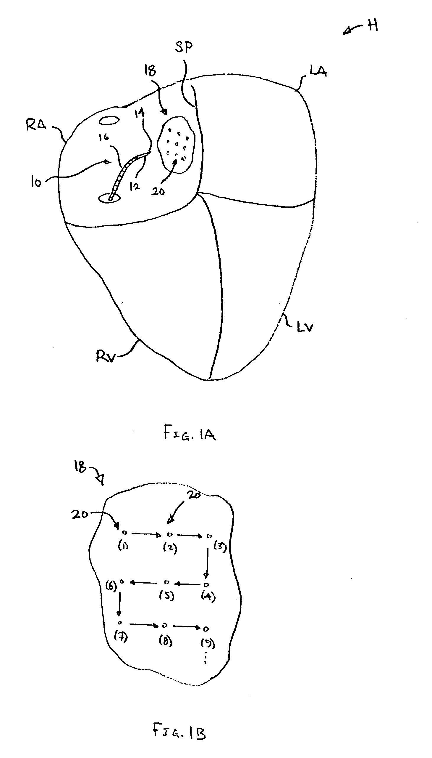
[0025] The left and right atria carry blood that varies in several physiological parameters. For example, the left atrium's blood oxygen saturation may reach 99% whereas the right atrium may only reach an oxygen saturation of 80% or lower. Another parameter includes pressure differences. The pressure in the left atrium is normally higher than the pressure in the right atrium, except in certain disease states. Also, the pressure wave forms have different amplitudes. Moreover, the wave forms also differ between the atria and other neighboring cardiac structures, such as the aorta (which has high velocity blood flow), and the intra-atrial septum or pericardium (which have no blood flow).

[0026] To gain access to the left atrium when performing a transeptal puncture, a clinician can perform a sounding procedure by utilizing any of the physiological parameter differences to detect a direct pathway. One method may be to utilize the oxygen saturation difference between the atrial chambers ...


PUM


Abstract

Left atrial access apparatus and methods are described herein. Different parameters, such as oxygen saturation difference, between the left and right atrial chambers is utilized to guide a needle or catheter into a desired position within the heart. Various sensing elements can be utilized to detect the physiological parameter difference, such as oxygen levels, in the left atrium. The sensor can be carried by the needle, at its tip or along its body, and can measure the physiological parameter levels contained in the blood, fluid, or tissue.

Description

CROSS-REFERENCE TO RELATED APPLICATIONS [0001] This application claims the benefit of priority to U.S. Provisional Pat. App. Ser. No. 60 / 506,465 filed Sep. 26, 2003, which is incorporated herein by reference in its entirety.FIELD OF THE INVENTION [0002] The present invention relates generally to medical devices used for detecting parameters within the body. More particularly, the present invention relates to apparatus and methods for facilitating or enabling access across the atrial septum, e.g., for septostomy procedures within the heart. BACKGROUND OF THE INVENTION [0003] The mammalian heart is divided into four chambers. The superior or upper two chambers include the left and right atria. The right atrium is fluidly in communication with the venous system and carries deoxygenated blood. The left atrium receives oxygenated blood from the lungs and facilitates the movement of this blood into the left ventricle for pumping throughout the body. [0004] The inferior vena cava (IVC) and...

Claims


Application Information

Patent Timeline
Patent Type & Authority Applications(United States)
IPC IPC(8): A61B5/00A61FA61M25/09
CPCA61B5/14542A61B5/6848A61B5/1459
Inventor SAADAT, VAHID
Owner HANSEN MEDICAL INC
Why Eureka
Try Eureka