

Method for fixing tissue
a tissue and tissue technology, applied in the field of tissue fixation, can solve the problems of thrombosis and platelet deposition, complete obstruction of the conduit, etc., and achieve the effects of promoting biocompatibility of biological tissue, reducing the formation of clots, and ensuring endothelization
- Summary
- Abstract
- Description
- Claims
- Application Information
AI Technical Summary
Benefits of technology
Problems solved by technology
Method used
Image

Examples
second embodiment
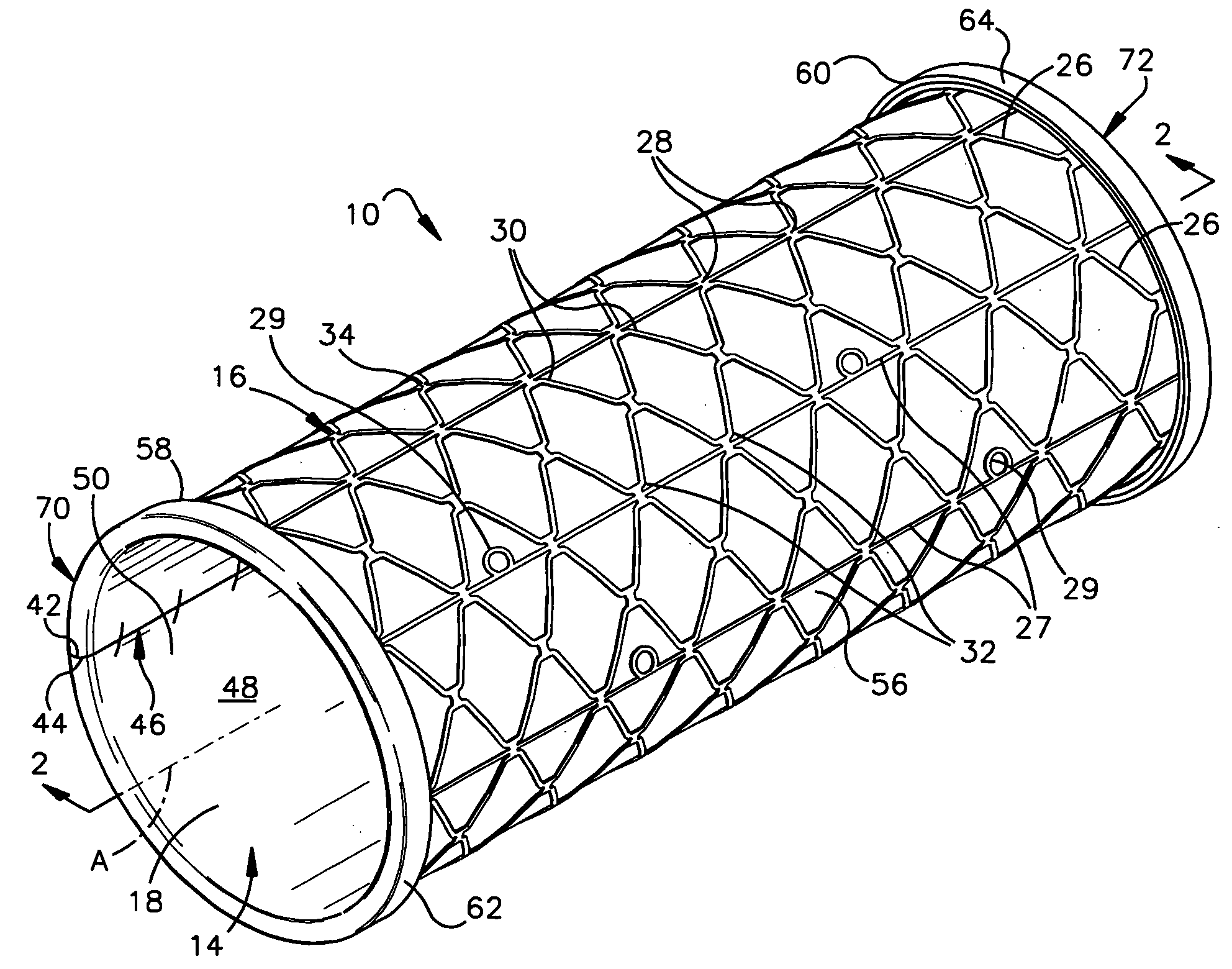
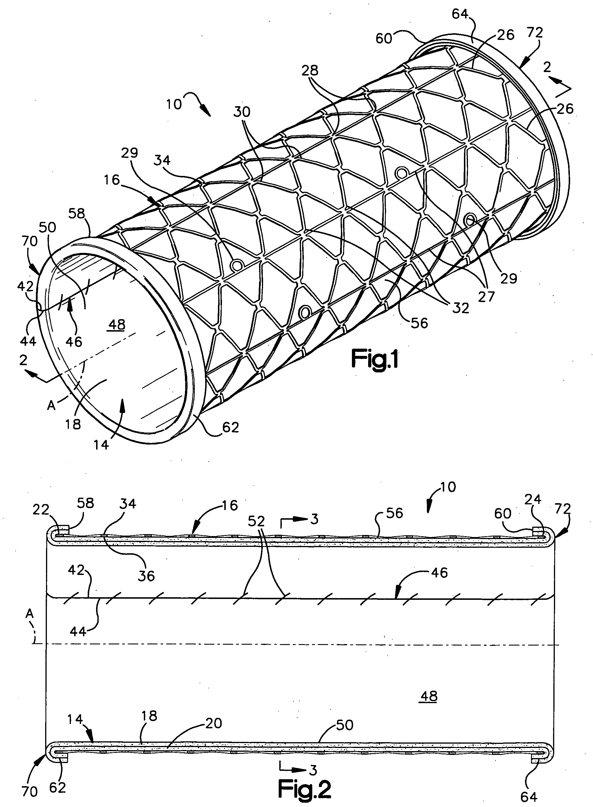
[0094]FIG. 6 is a longitudinal sectional view of an apparatus 10a constructed in accordance with the present invention. Structures of the embodiment shown in FIG. 6 that are similar to structures of FIGS. 1-3 have the same reference numbers with the suffix “a” added. The apparatus 10a is identical to apparatus 10 of FIGS. 1-3 with the exception that the layer of biological tissue 14a in the embodiment of FIG. 6 includes only a layer 18a of serous membrane.
[0095] The layer of biological tissue 14a is harvested to include only the layer 18a of serous membrane. The method for harvesting only a layer 18a of serous membrane is known in the art.
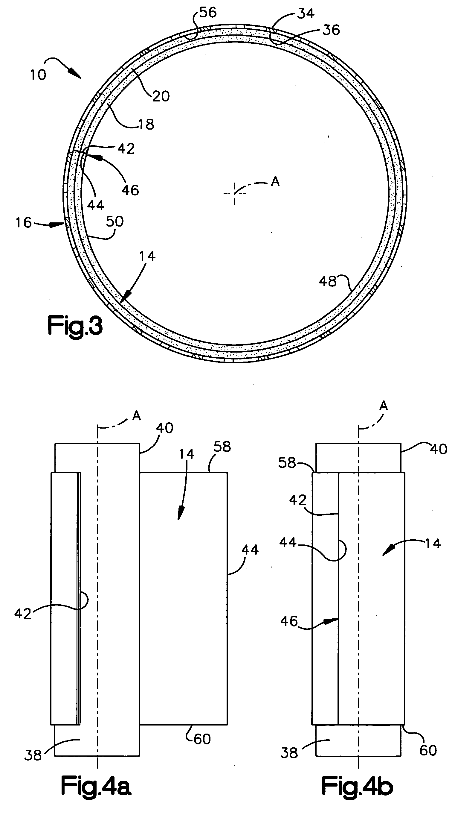
[0096] The assembly of apparatus 10a is identical to the assembly of apparatus 10 that is illustrated in FIGS. 4a-4f. When trimmed into the desired shape, the layer of biological tissue 14a includes first and second side surfaces 42a and 44a, respectively, and first and second axial ends 58a and 60a, respectively.
[0097] The assembled apparatus in...
third embodiment
[0099]FIG. 7 is a longitudinal sectional view of an apparatus 10b constructed in accordance with the present invention. Structures of the embodiment shown in FIG. 7 that are similar to structures of FIGS. 1-3 have the same reference numbers with the suffix “b” added.
[0100] The apparatus 10b illustrated in FIG. 7 includes a layer of biological tissue 14b and an expandable support member 16b. The layer of biological tissue 14b includes a serous membrane lining 18b and associated fascia lining 20b. The expandable support member 16b has a structure similar to that illustrated in FIG. 1. The layer of biological tissue 14b forms the innermost component of the apparatus 10b.
[0101] The layer is biological tissue 14b is formed into a tubular portion by abutting first and second side surfaces 42b and 44b of the biological tissue 14b at a seam 46b. Preferably, the first and second side surfaces 42b and 44b are sutured together at the seam 46b and biological glue (not shown) is applied to an o...
fourth embodiment
[0104]FIG. 8 is a longitudinal sectional view of an apparatus 10c constructed in accordance with the present invention. Structures of the embodiment shown in FIG. 8 that are similar to structures of FIG. 7 have the same reference numbers with the suffix “c” replacing the suffix “b”. The apparatus 10c is identical to apparatus 10b of FIG. 7 with the exception that the layer of biological tissue 14c in the embodiment of FIG. 8 includes only a layer 18c of serous membrane.
[0105] The assembly of apparatus 10c is identical to the assembly of apparatus 10b. When trimmed into the desired shape, the layer of biological tissue 14c includes first and second side surfaces 42c and 44c, respectively, and first and second axial ends 58c and 60c, respectively.
[0106] The assembled apparatus includes a seam 46c that is formed from abutting the first and second side surfaces 42c and 44c. The inner surface 48c of the assembled apparatus 10c is defined by the inner surface 50c of the layer 18c of sero...
PUM
| Property | Measurement | Unit |
|---|---|---|
| molar concentration | aaaaa | aaaaa |
| thickness | aaaaa | aaaaa |
| length | aaaaa | aaaaa |
Abstract
Description
Claims
Application Information