Medium for contrast enhancement or convenience for ultrasonic, endoscopic, and other medical examinations

a technology of contrast enhancement and ultrasonic, which is applied in the field of medium for contrast enhancement or convenience for ultrasonic, endoscopic, and other medical examinations, can solve the problems of inconvenient and messy fluid puddles in the examination area, inconvenient use of intrauterine infusion of nacl or other solution as ultrasound contrast enhancer, and further source of discomfort for patients, etc., to achieve convenient use, less pressure, and constant infusion

Active Publication Date: 2005-08-04
ULTRAST INC
View PDF4 Cites 17 Cited by
  • Summary
  • Abstract
  • Description
  • Claims
  • Application Information

AI Technical Summary

Benefits of technology

[0038] The medium of the invention distends the body or organ cavity with less pressure, and allows medical imaging of the cavity without concomitant, constant infusion of additional medium. Good quality vision typically is possible until the medium liquefies, at which time the examination may deteriorate due t...

Problems solved by technology

While simple in principle, the use of an intrauterine infusion of a NaCl or other solution as an ultrasound contrast enhancer is complex and cumbersome in practice.
The need to constantly flush contrast fluid through the uterine cavity while conducting the ultrasound examination, as it continually leaks out, also results in inconvenient and messy fluid puddles in the examination area, including on the examination table—which can be a further source of discomfort for the patient.
Needless to say, sonohysterography is considered a time-consuming procedure by most gynecologists, who tend to use it reluctantly and only in certain cases that are deemed to warrant the extra inconvenience and discomfort.
Despite these practical difficulties, the quality enhancement of ultrasound images achieved through sonohysterography is significant and allows more precise diagnoses of uterine pathologies, such as uterine fibroids and polyps, otherwise impossible or difficult to examine with plain ultrasounds.
Yet despite ample clinical value documented by many academic studies, actual use of sonohysterography remains relatively limited because of the difficulties and time-consuming nature of the procedure.
Irrespective of its indications, positive contrast sonohysterography is as cumbe...

Method used


Image

  • Medium for contrast enhancement or convenience for ultrasonic, endoscopic, and other medical examinations
  • Medium for contrast enhancement or convenience for ultrasonic, endoscopic, and other medical examinations
  • Medium for contrast enhancement or convenience for ultrasonic, endoscopic, and other medical examinations

Examples


example 1

Contrast Ultrasound —53 Patients

[0080] Design—Prospective pilot feasibility study in infertile women and women with dysfunctional uterine bleeding (DUB).

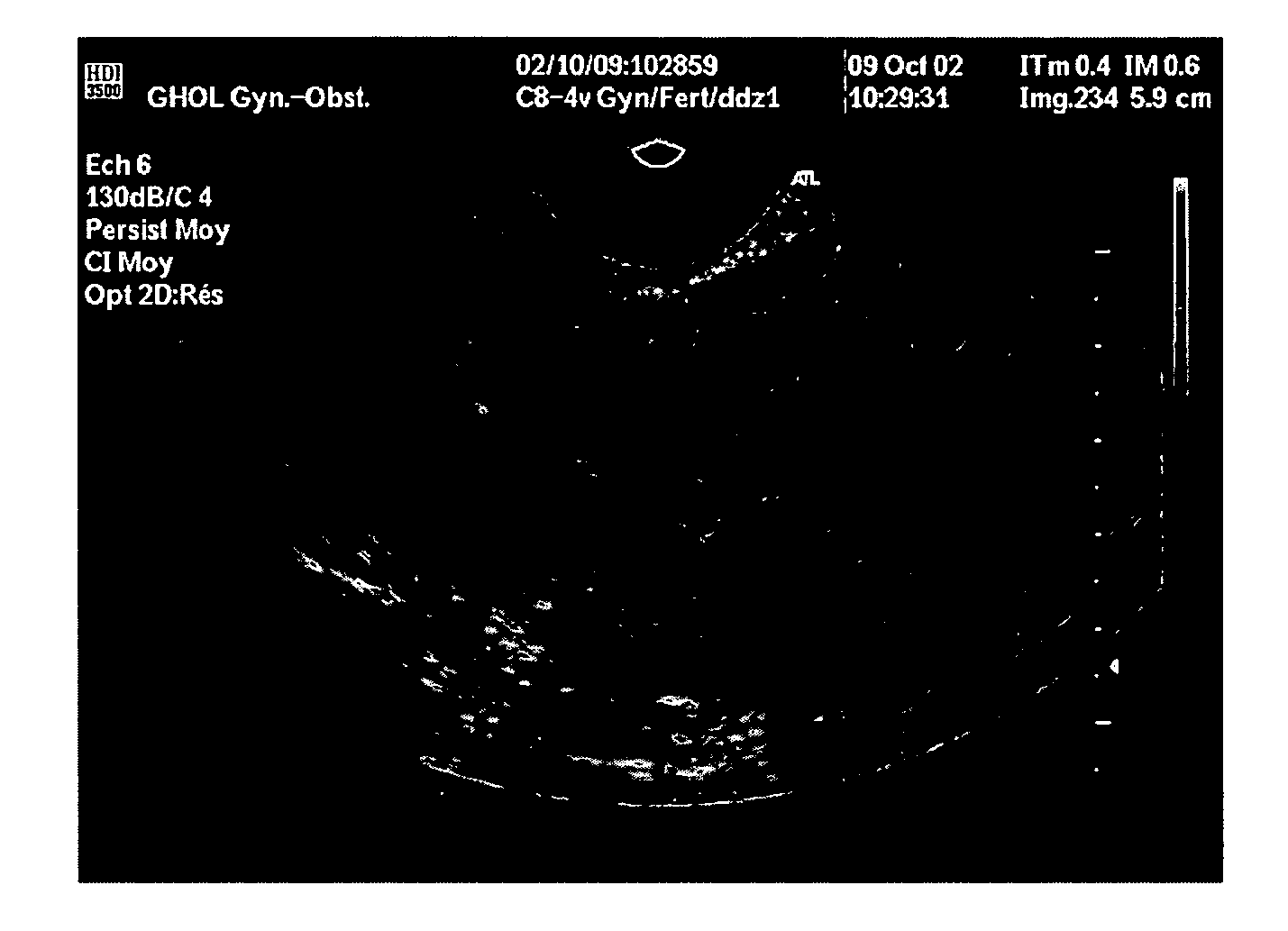
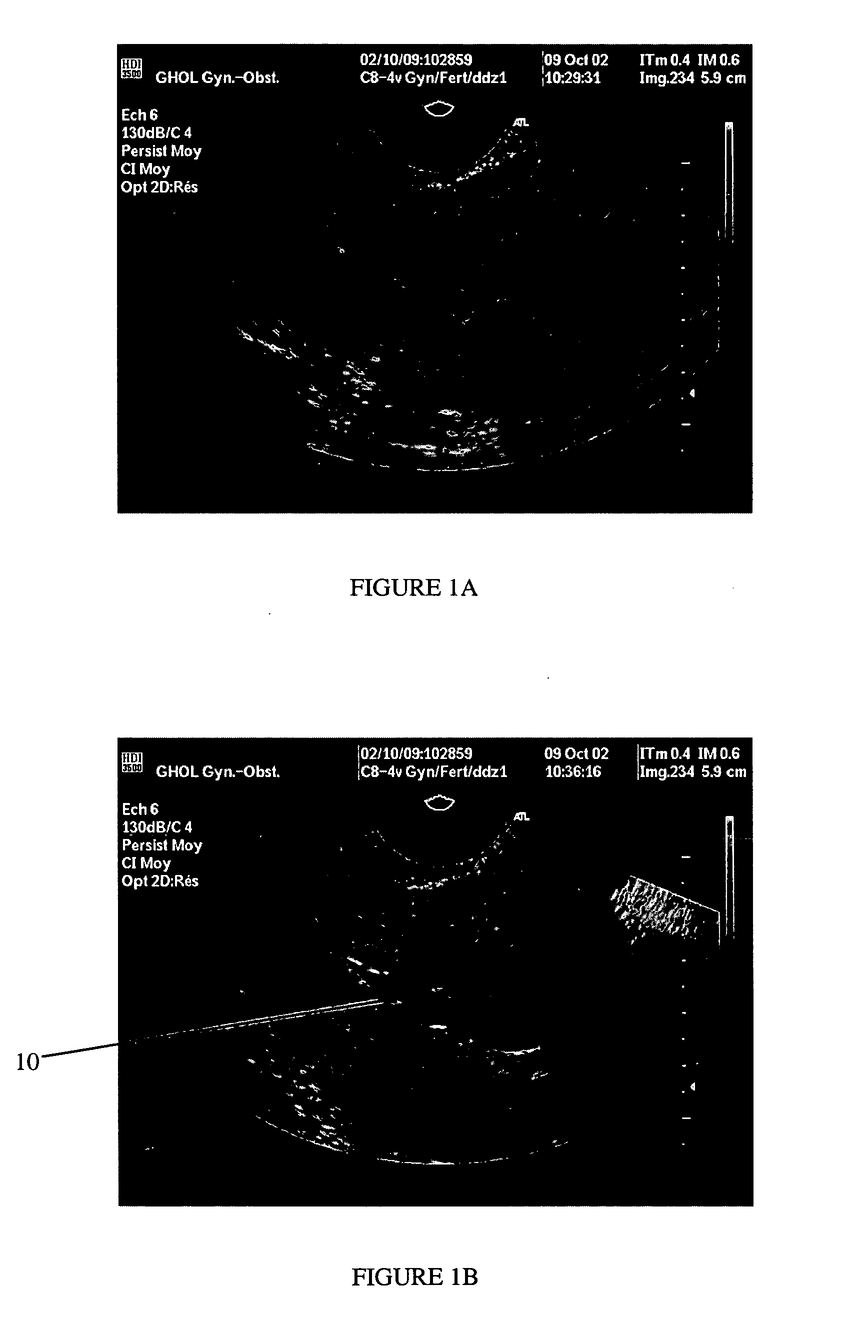
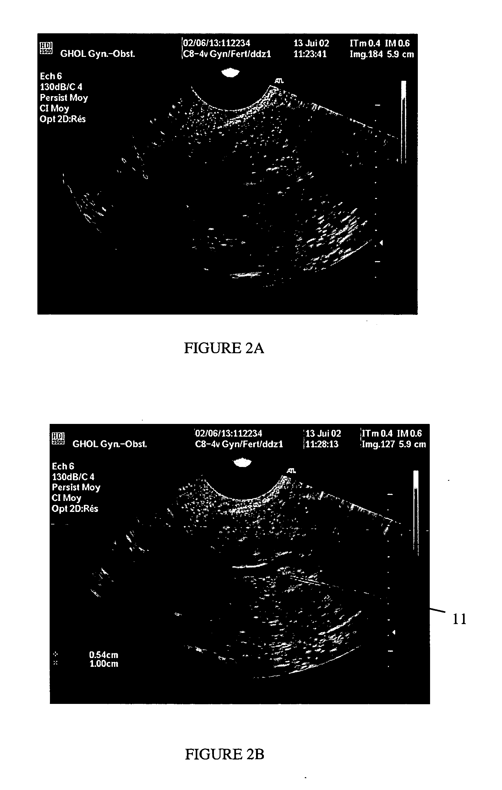
[0081] Materials and Methods —53 women consulting for infertility (n=27) or DUB (n=26) were studied. After a conventional vaginal ultrasound was performed, all underwent contrast ultrasound examination. For this, 3-7 cc of a negative (black) phase shifting uterine contrast medium (“PSCM”) was infused in the uterine cavity using a 3.5 Frydman embryo transfer catheter. The uterine catheter was then removed and another vaginal ultrasound was performed.

[0082] Results—In all women, the uterine cavity remained distended for ≧3 minutes, leaving a ≧3.5 mm thick “black” interface in the uterine cavity. In all patients uterine distension and medium had disappeared after 10 minutes. In 21 of 26 women presenting with DUB, contrast ultrasound revealed an intrauterine pathology that was subsequently confirmed by hysteroscopy and histology. In ...

example 2

Endoscopic Vision of the Uterine Cavity —7 Patients

[0085] Design—Pilot feasibility study in women scheduled for surgical hysteroscopy, using optically-transparent phase shifting medium (“PSM”).

[0086] Materials and Methods—The PSM was tested in 7 women immediately before a scheduled surgical hysteroscopy. 7-10 cc of PSM was infused in the uterine cavity using a common embryo transfer catheter after patients underwent cervical dilatation. After the embryo transfer catheter was removed, the conventional hysteroscope was introduced and endoscopic exploration undertaken without connecting the infusion system. After direct visualization was completed, conventional hysteroscopy was conducted using an infusion of a distending solution.

[0087] Results—The uterine cavity, slightly distended by the viscous nature of the PSM at the time of insertion, was easily explored with panoramic endoscopy. The viscous nature of the PSM also prevented it from mixing with blood originating from the uterin...

example 3

Positive and Negative Contrast Uterine Gels

[0091] A negative contrast gel would be particularly appropriate for use to enhance the contrast of uterine ultrasounds. Such a negative medium would facilitate the diagnosis of uterine pathologies such as sub-mucosal fibroids, endometrial polyps, endometrial synechiae (scars), uterine malformations and all uterine pathologies having endometrial repercussion and / or ultrasound expression. Potentially, the negative gel can permit imaging part or all of one or both Fallopian tubes.

[0092] Uterine exposure to progesterone or progestins, such as with oral contraception, is likely to prolong the presence of the gel because of the utero-relaxing properties of progesterone and progestins. And other possible applications will be sought for enhancing negative contrast of other forms of imaging of the uterus and tubes, including for example by MRI and CT scans.

[0093] Positive contrast media will also be useful. A positive contrast formulation likely...


PUM


Abstract

The present invention relates to a particularly convenient medium for providing contrast enhancement and/or distension of the subject body or organ cavity during imaging, radiographic, visualization, or other similar medical examinations, including ultrasound, endoscopic examinations, MRI, x-ray, hystero-salpingograms, CT scans, and similar procedures. The present invention provides the contrast enhancement and/or distends the body or organ cavity without constant leakage of the medium or the resulting need to constantly or repeatedly infuse additional medium. The medium is designed to have sufficient viscosity or consistence initially to remain in the body or organ cavity for a time generally sufficient to complete the procedure, and then to facilitate easy removal or expulsion from the cavity, preferably by liquefying or losing viscosity. The phase or viscosity change may be triggered by a variety of factors that liquefy or otherwise facilitate easy removal or expulsion of the composition from the cavity.

Description

CROSS-REFERENCE TO RELATED APPLICATIONS [0001] This application is a continuation of co-pending International Application No. PCT / EP03 / 04834, filed on May 8, 2003, which claims the benefit of U.S. Provisional Application No. 60 / 378,640, filed on May 9, 2002, and U.S. Provisional Application No. 60 / 428,145, filed on Nov. 18, 2002, the entire disclosures of which are hereby incorporated by reference herein.FIELD OF INVENTION [0002] The present invention relates to a particularly convenient medium for providing contrast enhancement or distension during imaging, radiographic, visualization, or other similar medical examinations and procedures involving a body or organ “virtual” cavity, including ultrasound, endoscopic examinations, MRI, x-ray, CT scans, and similar procedures jointly, “medical imaging procedures”). The present invention provides the contrast enhancement and / or distends a body or organ cavity that is normally virtual, without constant leakage of the medium or the need to...

Claims


Application Information

Patent Timeline
IPC IPC(8): A61BA61B1/00A61B5/05A61B5/055A61B8/02A61B5/00A61B8/06A61B8/12A61B8/14A61K49/00A61K49/22
CPCA61K49/226A61B8/00
Inventor DE ZIEGLER, DOMINIQUE
Owner ULTRAST INC
Why Eureka
Try Eureka