Nuclear medical diagnostic equipment and data acquisition method for nuclear medical diagnosis

a technology of nuclear medical diagnosis and data acquisition, which is applied in the field of nuclear medical diagnostic equipment and data acquisition methods for nuclear medical diagnosis, can solve the problems of data not being acquired, whole data acquisition time period, image quality of acquired image, including positional resolution and contrast, and inevitably degrades, so as to achieve higher sensitivity and positional resolution. the effect of resolution

Active Publication Date: 2005-08-25
TOSHIBA MEDICAL SYST CORP
View PDF5 Cites 44 Cited by
  • Summary
  • Abstract
  • Description
  • Claims
  • Application Information

AI Technical Summary

Benefits of technology

[0021] Besides, in the data acquisition of nuclear medicine wherein data cannot possibly be acquired in one time of breath holding period, an image can be taken at a higher positional resolution and a higher sensitivity in compliance with clinical purposes, with the breath holding conducted in a state where the patient is hardly burdened.

Problems solved by technology

Therefore, the data cannot be acquired in the state of one time of breath holding by the patient, over the whole data acquisition time period.
Thus, the image quality of the acquired image, including a positional resolution and a contrast, degrades inevitably.
In the case of the above respiratory-gated data acquisition, however, acquisition counts per finer phase decrease basically, and hence, statistic noise and the like noise become predominant, to pose the problem that the degradation of an image is incurred due to the noise.
In such a case where the breathing state cannot be reliably sensed, the extension of the data acquisition time period or the decrease of the acquisition counts is incurred, resulting in a situation where the throughput of the patient lowers or where the image degradation becomes drastic.
Therefore, the respiratory-gated data acquisition is not a little influenced by the bodily motions ascribable to the respiration, as compared with data acquisition in the non-breathing state of the patient, so that the image quality of the acquired image is unsatisfactory.
Meanwhile, as another problem it is permitted to acquire the data only in the respiratory rest periods of the patient.
In this case, however, data are not acquired in the respiratory periods or breathing state of the patient.
This poses the problem that the number of samplings decreases to degrade the smoothness, namely, sensitivity of the image.

Method used


Image

  • Nuclear medical diagnostic equipment and data acquisition method for nuclear medical diagnosis
  • Nuclear medical diagnostic equipment and data acquisition method for nuclear medical diagnosis
  • Nuclear medical diagnostic equipment and data acquisition method for nuclear medical diagnosis

Examples


first embodiment

2. Modification to First Embodiment

[0067]FIG. 4 shows a modification to the first embodiment. A flow chart in FIG. 4 shows the process of the “intermittent data acquisition method” applied to the SPECT method as has expanded the process shown in FIG. 2. In FIG. 4, steps which execute processing identical or equivalent to the processing of the foregoing steps in FIG. 2 are denoted by the same signs.

[0068] In the “intermittent data acquisition method” applied to the SPECT method, the gamma camera 13 is first located in an initial radiographing direction (position) by the system controller 25 (step S1′), a command for the start of breath holding is given (step S2), and acquired data are recorded with identification information based on marking, for a time period t1 (step S3). Subsequently, a command for the interruption of the breath holding is given (step S4). Further, whether or not data acquisitions in the current radiographing direction (position) have been completed is judged (st...

second embodiment

4. Modification to Second Embodiment

[0085]FIG. 8 shows a modification to the second embodiment. A flow chart in FIG. 8 shows the process of the “intermittent data acquisition method” applied to a SPECT method as has expanded the process shown in FIG. 6. In FIG. 8, steps which execute processing identical or equivalent to the processing of the foregoing steps in FIG. 6 are denoted by the same signs.

[0086] In the “intermittent data acquisition method” applied to the SPECT method, the gamma camera 13 is first located in an initial radiographing direction (position) by the system controller 25 (step S21′), and the same processing steps as in FIG. 6 are thereafter executed (steps S22-S29). Further, whether or not data acquisitions in all radiographing directions (positions) have been completed is judged by the system controller 25 (step S30). In a case where the judgment becomes “NO”, the gamma camera 13 is spatially moved into the next radiographing direction (position), and similar da...

third embodiment

5. Third Embodiment

[0089] Next, the third embodiment of the invention will be described with reference to FIGS. 9-11.

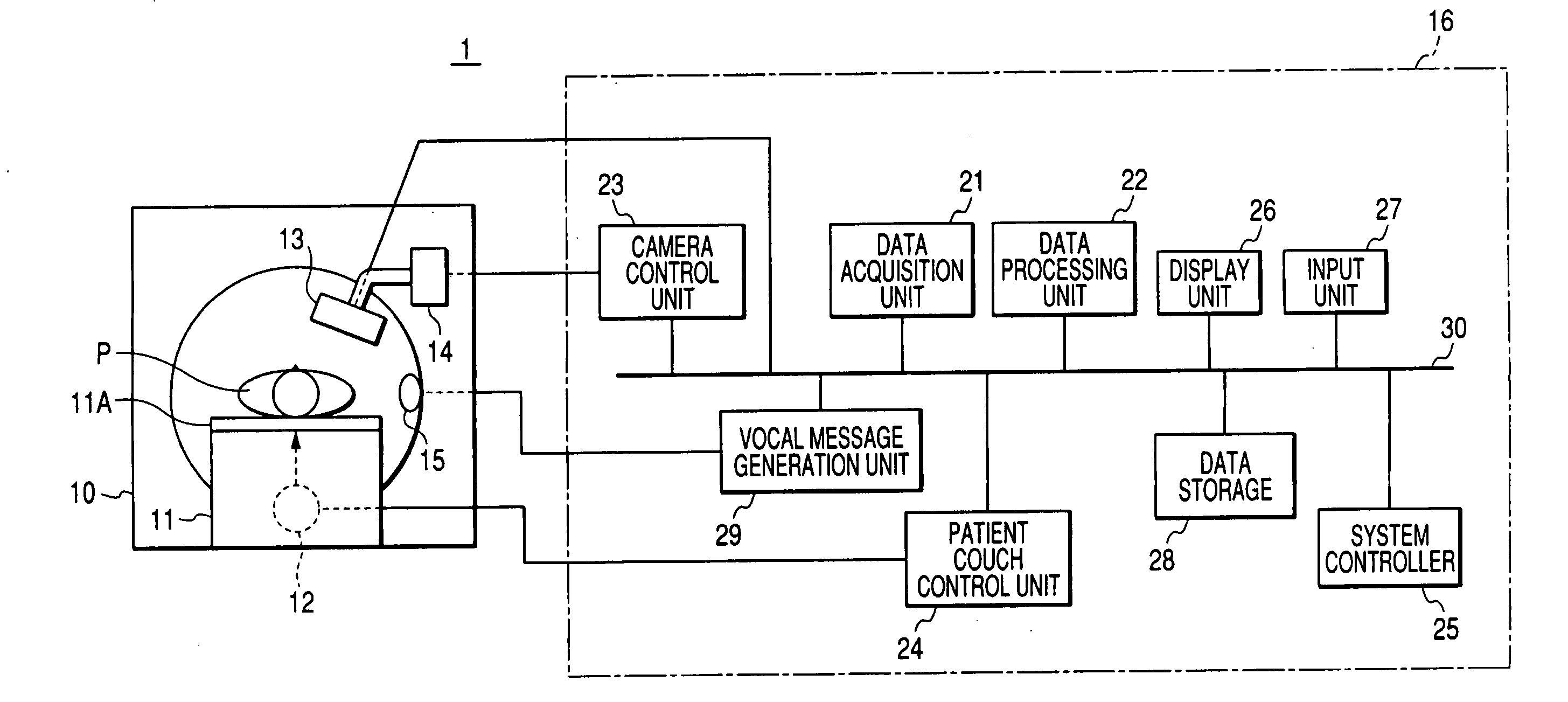
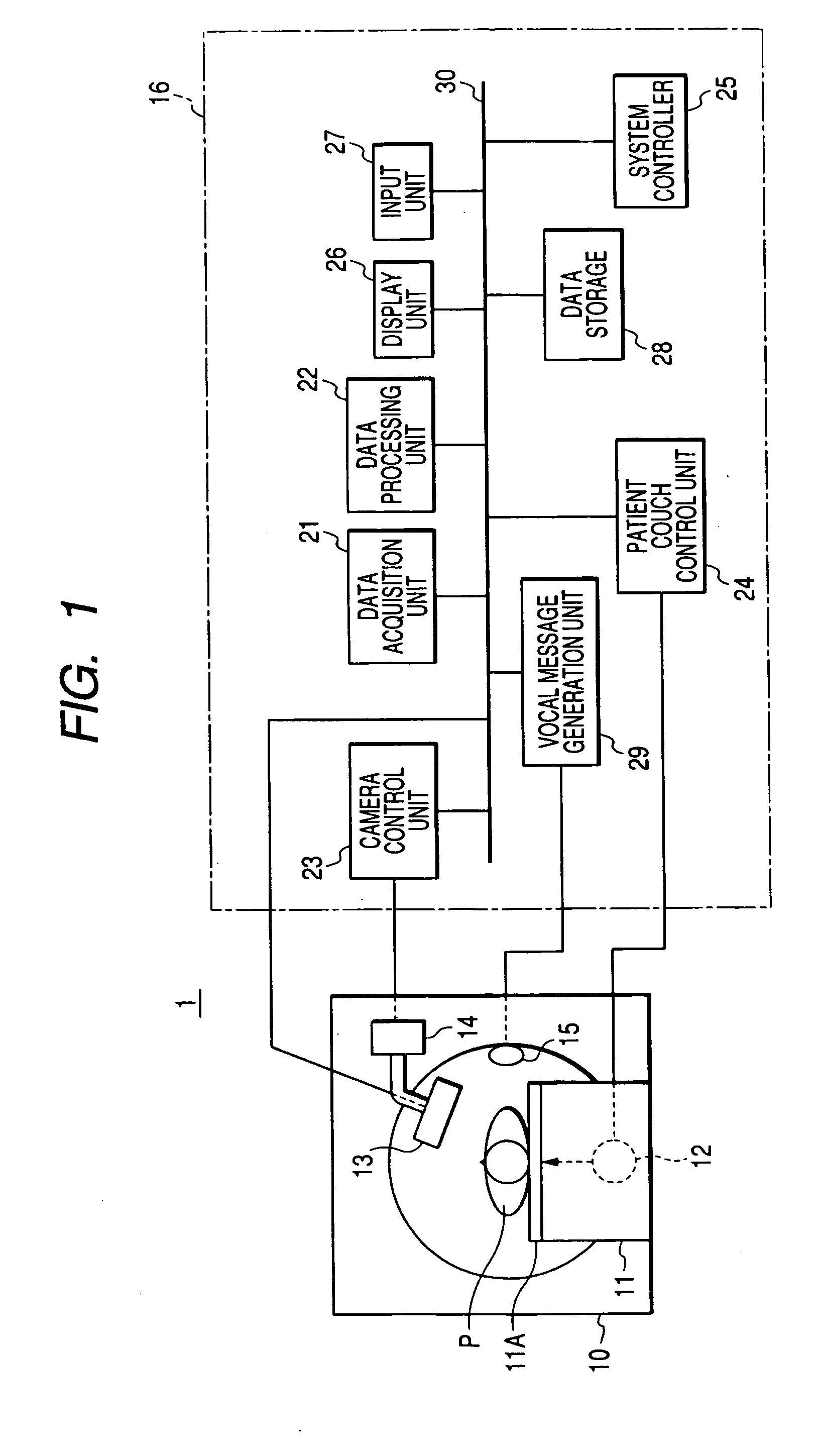
[0090] A nuclear medical diagnostic equipment according to the third embodiment concerns a construction which performs a SPECT method based on the “intermittent data acquisition method”. In particular, this embodiment features that, when a gamma camera 13 is rotated round a patient P stepwise so as to acquire data in each of a plurality of radiographing directions (positions), breathings and temporary breath stops based on the breath holdings are alternately iterated. On this occasion, the plurality of radiographing directions (positions) are fined so as to double those of the ordinary SPECT method in number (for example, 3 degrees per step, and 10-15 seconds in terms of an acquisition time period), and such acquisitions are iterated (for example, iterated 60 times). The outline of the process of this embodiment is shown in FIG. 9. Incidentally, the hardware architec...


PUM


Abstract

A nuclear medical diagnostic equipment wherein radiation which is emitted by a nuclide administered into the body of a patient is detected as projection data by a gamma camera, and an image which indicates the distribution of the nuclide within the body of the patient is obtained on the basis of the projection data. The equipment comprises a rotation unit which rotates the radiation detector round the patient, a respiration identification unit which identifies breathing of the patient and non-breathing thereof based on breath holding, a data storage unit in which the radiation detection data acquired by the radiation detector are stored in an identifiable manner on the basis of a result of the identification by the respiration identification unit, and an image generation unit which generates the image from the radiation detection data stored in the data storage unit on the basis of the result of the identification by the respiration identification unit.

Description

BACKGROUND OF THE INVENTION [0001] 1. Field of the Invention [0002] The present invention relates to a nuclear medical diagnostic equipment and a data acquisition method for a nuclear medical diagnosis wherein radiation emitted from a nuclide (radioisotope: RI) administered to a patient is detected using a gamma (γ) camera, and an RI distribution is imaged on the basis of the detection information. [0003] 2. Description of the Related Art [0004] With the technological advancements of hardware and software in recent years, a nuclear medical diagnostic equipment being an equipment for medical use has also made great progress. [0005] The nuclear medical diagnostic equipment includes a measurement unit called “gamma camera”, and it is employed for implementing the nuclear medical examination of a patient. The nuclear medical examination is implemented in such a way that, as stated above, a drug labeled with a radioisotope (hereinbelow, abbreviated to “RI”) is administered into the body ...

Claims


Application Information

Patent Timeline
Patent Type & Authority Applications(United States)
IPC IPC(8): G01T1/161A61B5/05A61B6/00G01T1/164G01T1/166G03B42/02
CPCA61B6/541A61B6/037
Inventor MOTOMURA, NOBUTOKUHAYASHI, MASUOHORIUCHI, SYOUJINISAWA, YOSHIFUMI
Owner TOSHIBA MEDICAL SYST CORP
Why Eureka
Try Eureka