Radiopaque, coaxial orthopedic tether design and method

a coaxial and radiopaque technology, applied in the field of orthopaque, can solve the problems of single cord not showing acceptable imaging characteristics under commonly used diagnostic imaging techniques, unable to effectively immobilize/or support the adjoining bone portions, and unable to achieve the effect of immobilizing the bone pieces of the joint, etc., and achieve the effect of improving image characteristics

Inactive Publication Date: 2005-09-01
WARSAW ORTHOPEDIC INC
View PDF37 Cites 176 Cited by
  • Summary
  • Abstract
  • Description
  • Claims
  • Application Information

AI Technical Summary

Benefits of technology

[0014] In other forms the present invention provides a surgical tether for orthopedic treatment to secure to two adjacent bone portions. The surgical tether comprises: a cord having a tensile strength sufficient to maintain a desired distance or orientation of the two bone portions; a first sheath substantially encasing the cord, wherein the first sheath comprises a plurality of fibers and provides an abrasion resistant coating to the cord; and means for imparting enhanced image characteristics to the tether.

Problems solved by technology

For articulating bone joints, such as the knees, hips and spinal column that have become damaged, bone plates alone may not be effective to either immobilize the bone pieces of the joint and / or support the adjoining bone portions.
The single cord does not exhibit acceptable imaging characteristics under commonly used diagnostic imaging techniques, i.e., x-ray, fluoroscopy, CT, and MRI imaging techniques.
Furthermore, for articulating joints the single cord can chafe against adjacent structures, whether those structures be adjacent bone structures or implanted devices such as bone plates, rods, screws, and the like.
The chafing is undesirable because it weakens the surgical cord by cutting either part-way or completely through one or more of the filaments making up the cord.
Furthermore, the frayed cord can irritate the surrounding tissue structure, which can be particularly painful for the patient.

Method used


Image

  • Radiopaque, coaxial orthopedic tether design and method
  • Radiopaque, coaxial orthopedic tether design and method
  • Radiopaque, coaxial orthopedic tether design and method

Examples


Embodiment Construction

[0018] For the purposes of promoting an understanding of the principles of the invention, reference will now be made to the embodiments illustrated herein, and specific language will be used to describe the same. It will nevertheless be understood that no limitation of the scope of the invention is thereby intended. Any alterations and further modifications in the described medical devices, surgical tethers, tether compositions, methods for treating patients, methods for preparing the devices, and any further applications of the principles of the invention as described herein are contemplated as would normally occur to one skilled in the art to which the invention relates.

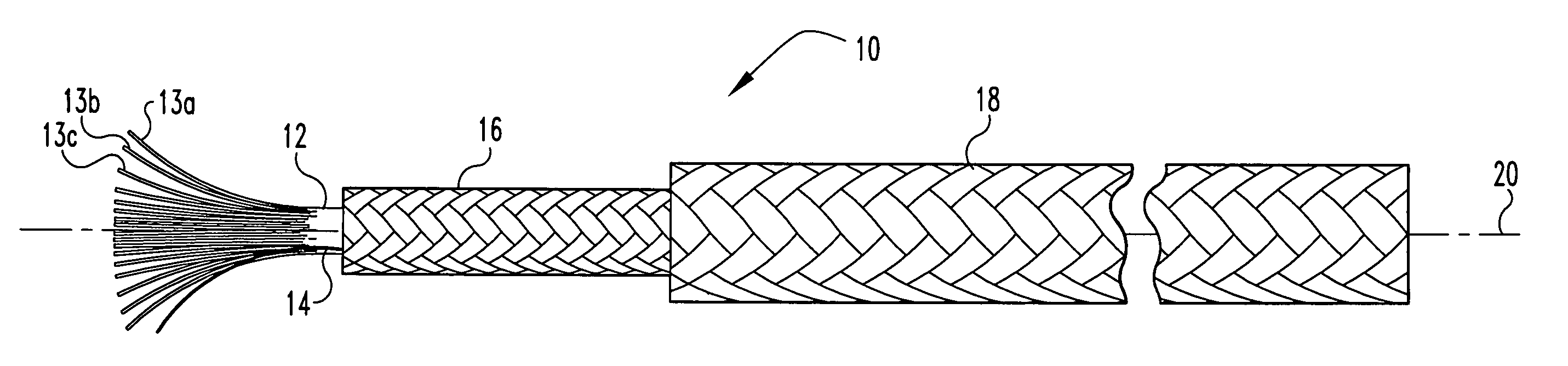
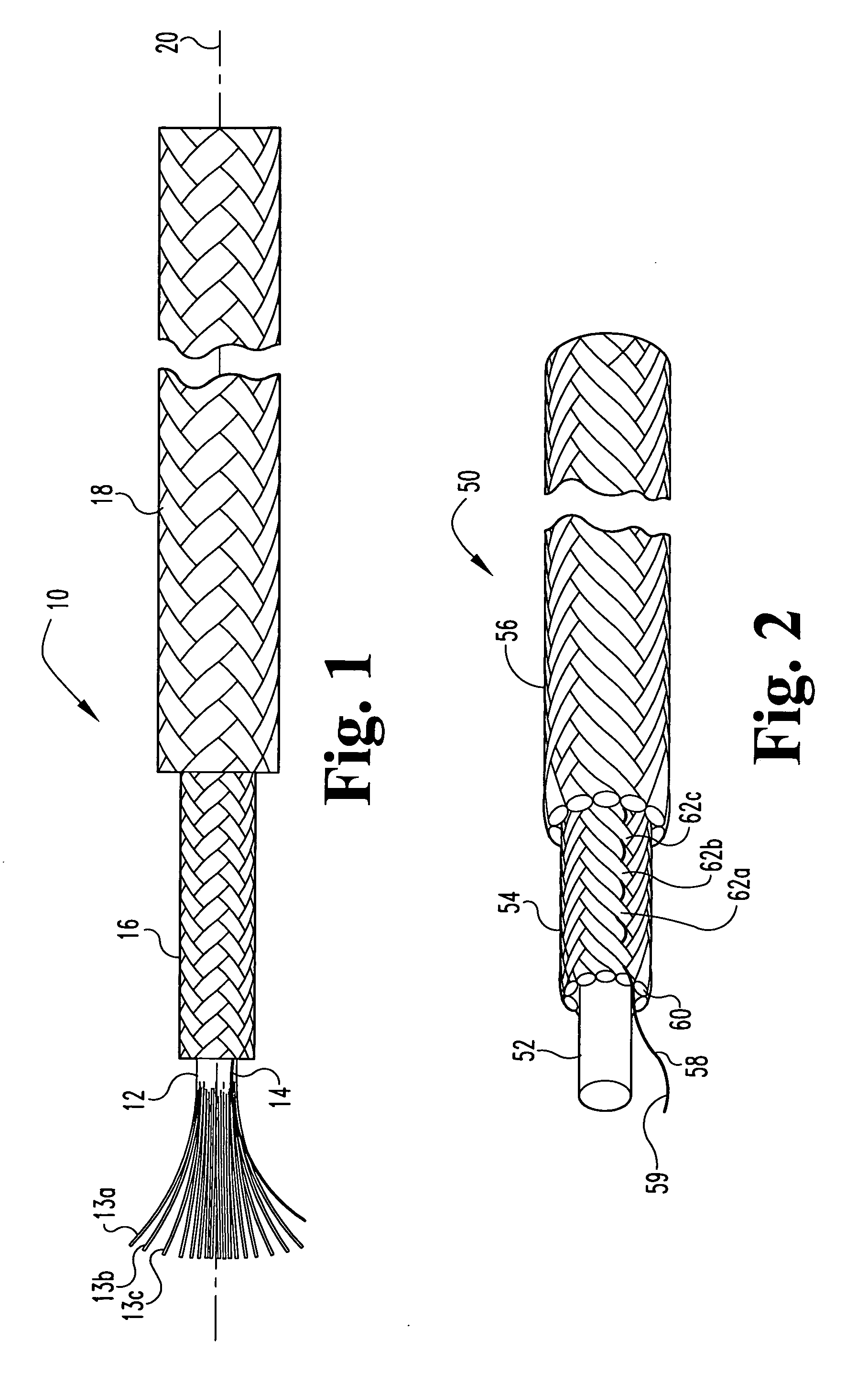
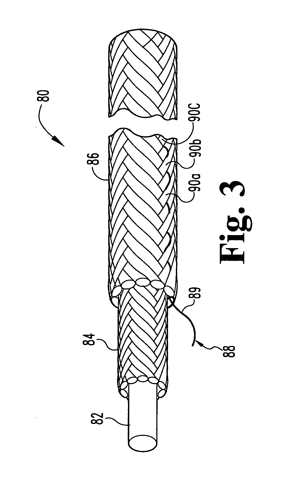
[0019] The present invention generally relates to a surgical device that includes an orthopedic tether that provides advantageous properties to treat bone defects. The device can be used to treat a variety of bone defects including diseased, damaged, and / or fractured bone. The defective bone structures can be the ...


PUM


Abstract

This invention relates to orthopedic tethers and their use to treat orthopedic defects. The tethers of the present invention include a central or inner load bearing fiber or cable surrounded by one or more protective sheaths. Additionally the tethers of the present invention provide enhanced imaging characteristics and can include one or more a radiopaque elements that can readily observed under common diagnostic imaging techniques.

Description

BACKGROUND OF THE INVENTION [0001] The present invention related to orthopedic devices for use in treating orthopedic defects. More specifically, the present invention is directed to orthopedic tethers to bind or secure bone and bone fragments together or to an ancillary orthopedic device; to methods of treating a patent with an orthopedic defect; and to methods of producing the orthopedic tether device. [0002] Orthopedic defects are frequently treated by joining or securing the damaged or diseased bone portions together thereby allowing the bone to heal. The bones can be partly or fully immobilized to promote bone tissue growth or regeneration and / or healing of stretched or torn ligaments. Immobilization and or joining of the bone pieces is usually accomplished using a variety of bone plates, surgical cord, and some type of fastening device such as a screw, staple, or glue. [0003] For articulating bone joints, such as the knees, hips and spinal column that have become damaged, bone...

Claims


Application Information

Patent Timeline
Patent Type & Authority Applications(United States)
IPC IPC(8): A61B17/00A61B17/68A61B17/70A61B17/84A61F2/00A61F2/08
CPCA61B17/68A61B17/70A61B17/842A61F2250/0098A61F2/08A61F2002/3008A61B2017/00004D04C1/12D10B2509/00
Inventor MOLZ, FRED J.DREWRY, TROY D.
Owner WARSAW ORTHOPEDIC INC
Why Eureka
Try Eureka