CTLA4-Cy4 fusion proteins

a technology of fusion proteins and ctla4cy4, which is applied in the field of fusion proteins, can solve the problems of undesirable side effects and detrimental effects in the subject, and achieve the effects of suppressing cell-mediated immune responses, reducing complement activation ability, and provoking antigen-specific t cell toleran

Inactive Publication Date: 2005-09-08
REPLIGEN CORP
View PDF7 Cites 24 Cited by
  • Summary
  • Abstract
  • Description
  • Claims
  • Application Information

AI Technical Summary

Problems solved by technology

Fc receptor-mediated phagocytosis or antibody-dependent cellular cytotoxicity, may induce detrimental side effects in the subject and are therefore undesirable.

Method used


Image

  • CTLA4-Cy4 fusion proteins
  • CTLA4-Cy4 fusion proteins
  • CTLA4-Cy4 fusion proteins

Examples


example 1

Preparation of CTLA4-Immunoglobulin Fusion Proteins with Reduced Effector Function

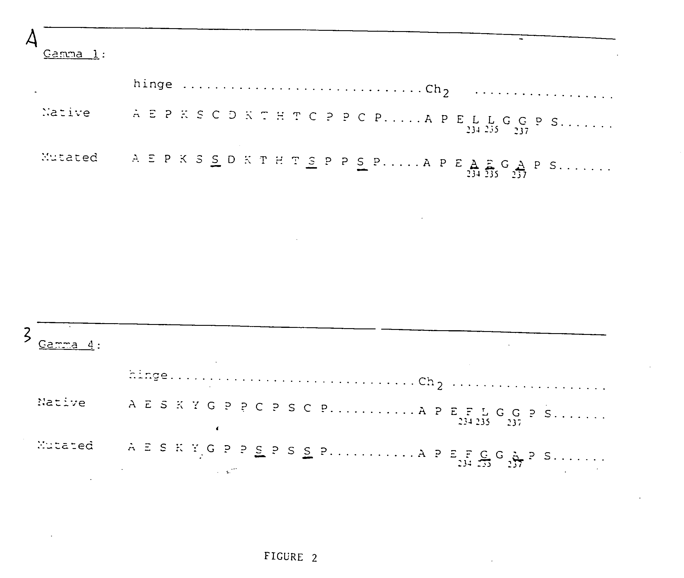
[0113] The extracellular portion of the T cell surface receptor CTLA4 was prepared as a fusion protein coupled to an immunoglobulin constant region. The immunoglobulin constant region was genetically modified to reduce or eliminate effector activity inherent in the immunoglobulin structure. Briefly, DNA encoding the extracellular portion of CTLA4 was joined to DNA encoding the hinge, CH2 and CH3 regions of human IgCγ1 or IgCγ4 modified by directed mutagenesis. This was accomplished as follows:

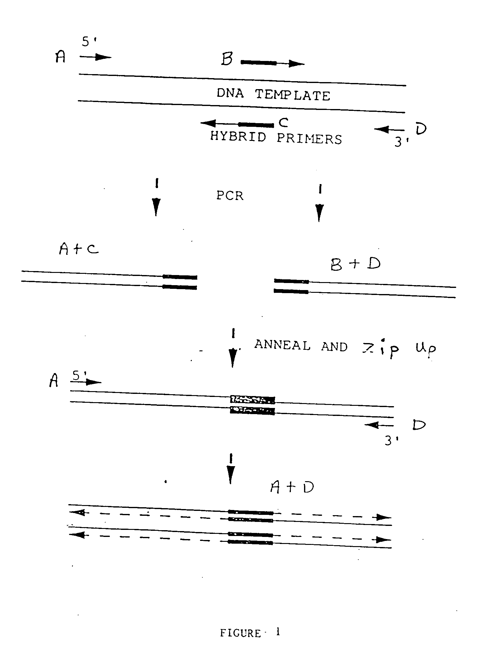
Preparation of Gene Fusions

[0114] DNA fragments corresponding to the DNA sequences of interest were prepared by polymerase chain reaction (PCR) using primer pairs described below. In general, PCR reactions were prepared in 100 μl final volume composed of Taq polymerase buffer (Gene Amp PCR Kit, Perkin-Elmer / Cetus, Norwalk, Conn.) containing primers (1 μM each). dNTPs (200 μM each), 1 ng of template DNA, and Ta...

example 2

Characterization of CTLA4 Fusion Proteins

[0143] The ability of the various CTLA4-Ig forms and CTLA4Ab to bind to their counter receptors B7-1 (Freeman, G. F., et al. (1988) J. Immunol. 143:2714-2722) and B7-2 (Freeman, G. F., et al., (1993) Science 262: 909-911) was demonstrated using the following assays.

A. Fluorescence Activated Cell Staining (FACS).

[0144] Purified preparations of the various recombinant CTLA4 forms were tested for their ability to bind to transfected COS cell transiently expressing hB7-1 or hB7-2 or transfected CHO cells stably expressing hB7-1 or hB7-2. The recombinant CTLA4 protein (10 μg / ml) was incubated with B7 expressing cells (2×10⁶ cells) for 1 hr on ice in FACS wash solution (1% bovine serum albumin in PBS). The cells were washed 3 times with FACS wash solution. The cell bound CTLA4 was detected by reaction with anti-human Ig-FITC (Dako Corporation, Carpintera, Calif.) or protein A-FITC (Dako) for 30 mintues on ice in the dark. The cells were washed ...

example 3

Preparation of E. coli-Expressed Human CTLA4

A. Intracellular Expression of CTLA4 in E. coli

1. Cloning and Expression of CTLA4 Extracellular Domain

[0160] The extracellular domain of CTLA4 was expressed in E. coli after cloning into expression vector pETCm11a. This vector was derived from expression vector pET-11a (Novagen Inc., Madison Wis.) by cloning a chloramphenicol resistance gene cassette into the ScaI restriction site within the ampicillin resistance gene. The extracellular domain of CTLA4 was prepared from plasmid phCTLA4 by PCR amplification using oligonucleotide 5′GCAGAGAGACAT ATGGCAATGCACGTGGCCCAGCCTGCTGTGG-3′ (SEQ ID NO: 20) as forward primer and oligonucleotide 5′-GCAGAGAGAGGATCCTCAGTCAGTTAGT CAGAATCTGGGCACGGTTCTGG-3′ (SEQID NO: 21) as reverse primer. The forward PCR primer (SEQ ID NO: 20) contains an NdeI restriction site in which the ATG sequence in the NdeI restriction site is followed immediately by the codon for the first amino acid of mature CTLA4 (Dariavach, ...


PUM

PropertyMeasurementUnit
volumeaaaaaaaaaa
concentrationaaaaaaaaaa
pHaaaaaaaaaa

Abstract

CTLA4-immunoglobulin fusion proteins having modified immunoglobulin constant region-mediated effector functions, and nucleic acids encoding the fusion proteins, are described. The CTLA4-immunoglobulin fusion proteins comprise two components: a first peptide having a CTLA4 activity and a second peptide comprising an immunoglobulin constant region which is modified to reduce at least one constant region-mediated biological effector function relative to a CTLA4-IgG1 fusion protein. The nucleic acids of the invention can be integrated into various expression vectors, which in turn can direct the synthesis of the corresponding proteins in a variety of hosts, particularly eukaryotic cells. The CTLA4-immunoglobulin fusion proteins described herein can be administered to a subject to inhibit an interaction between a CTLA4 ligand (e.g., B7-1 and / or B7-2) on an antigen presenting cell and a receptor for the CTLA4 ligand (e.g., CD28 and / or CTLA4) on the surface of T cells to thereby suppress an immune response in the subject, for example to inhibit transplantation rejection, graft versus host disease or autoimmune responses.

Description

BACKGROUND OF THE INVENTION [0001] To induce antigen-specific T cell activation and clonal expansion, two signals provided by antigen-presenting cells (APCs) must be delivered to the surface of resting T lymphocytes (Jenkins, M. and Schwartz, R. (1987) J. Exp. Med. 165:302-319; Mueller, D. L., et al. (1990) J. Immmunol. 144:3701-3709; Williams, I. R. and Unanue, E. R. (1990) J. Immunol. 145:85-93). The first signal, which confers specificity to the immune response, is mediated via the T cell receptor (TCR) following recognition of foreign antigenic peptide presented in the context of the major histocompatibility complex (MHC). The second signal, termed costimulation, induces T cells to proliferate and become functional (Schwartz, R. H. (1990) Science 248:1349-1356). Costimulation is neither antigen-specific, nor MHC restricted and is thought to be provided by one or more distinct cell surface molecules expressed by APCs (Jenkins, M. K., et al. (1988) J. Immunol. 140:3324-3330; Linsl...

Claims


Application Information

Patent Timeline
Patent Type & Authority Applications(United States)
IPC IPC(8): A61K38/00C07K14/705C12N1/21
CPCA61K38/00C07K2319/00C07K14/70503A61K2039/5154A61P37/06
Inventor GRAY, GARYCARSON, JERRYJAVAHERIAN, KASHIRENNERT, PAULSILVER, SANDRA
Owner REPLIGEN CORP
Why Eureka
Try Eureka