Combination of transplantation and oncolytic virus treatment

a technology of oncolytic virus and transplantation, which is applied in the field of virus pretreatment, can solve the problems of unable to achieve 100% success in depleting tumor cells in clinical setting, and unable to achieve 100% success in reducing the patient's resistance to viral infections, etc., to achieve the effect of assessing the cytopathic

Inactive Publication Date: 2005-09-29
ONCOLYTICS BIOTECH
View PDF9 Cites 23 Cited by
  • Summary
  • Abstract
  • Description
  • Claims
  • Application Information

AI Technical Summary

Benefits of technology

[0220] Cells lines grown to subconfluence and purified or enriched primary tumor samples in culture media were infected with reovirus at a multiplicity of infection (MOI) of 40 plaque-forming units (PFU) / cell. To assess cytopathic effect, cells were photographed under a light microscope at 48 or 72 hours after infection.
[0221] Cell lines (U937 and RPMI 8226) were grown to subconfluence and infected with reovirus at an MOI of 40 PFU / cell. To evaluate whether reovirus infects AP cells, cells were cultured in RPMI medium containing 10% FBS in the presence or absence of granulocyte colony-stimulating factor (G-CSF) (10 ng / mL) and infected with reovirus at 40 MOI. At various time points after infection, the medium was replaced with media containing 0.1 μCi / mL (0.0037 MBq / mL) (35S)-methionine. After further incubation for 12 hours at 37° C., the cells were washed in PBS and lysed in lysis buffer containing 1% Triton X-100, 0.5% sodium deoxycholate, and 1 mM EDTA (ethylenediaminetetraacetic acid). The nuclei were then removed by low-speed centrifugation, and the supernatants were stored at −80° C. until use. Radiolabeled lysates were subjected to sodium dodecyl sulfate-polyacrylamide gel electrophoresis (SDS-PAGE) as previously described (Lee, et al. 1981).
[0222] Monocytic (U937) and myeloma (RPMI 8266) cells were cultured in the presence or absence of live virus (40 MOI) for up to 7 days. At 0, 1, 2, 3, 4, and 7 days after virus infection, cells were harvested, and 1 mL cell culture suspension containing approximately 1×10⁶ cells was centrifuged at 750g for 1 minute. The cell pellet was resuspended in 1 mL of 50 μg / mL propidium iodide / RNase / Triton X-100 (Sigma Chemical, St Louis, Mo.), and 100 μL Flow-count beads (Beckman Coulter, Hialeah, Fla.) was added to each tube. Intact cells were enumerated using flow-count beads as an internal calibrator.
[0223] CD34-phycoerythrin (PE) (581) and CD45-fluorescein isothiocyanate (FITC) (J33) (Beckman-Coulter) antibodies were added to 100 μL diluted AP cells using a reverse pipetting technique to ensure accuracy. Samples were incubated for 10 minutes at room-temperature in the dark. Flow-count beads (100 μL) were added to each tube using the same technique as in “Cell counting using flow cytometry.” Flow cytometric analysis was performed on an EPICS XL flow cytometer (Beckman Coulter) using a modified ISHAGE strategy (Keeney, et al. 1998; Sutherland, et al. 1996). Data from 4 parameters were collected for analysis: forward scatter (FS), log side scatter (LSS), log fluorescence 1 (LFL1), and log fluorescence 2 (LFL2). Acquisition was halted at 100,000 CD45+ events. Hematopoietic progenitor cells were identified and counted in 2 histograms (CD45-FITC versus CD34-PE and FS versus LSS) using ISHAGE criteria: dim CD45+, bright CD34+ form a discrete cell cluster with a larger FS signal than lymphocytes. The use of a known amount of Flow-Count fluorospheres allowed the determination of absolute CD34+ cell count directly from the flow cytometer.
[0224] Apheresis product cells were depleted of lineage-committed cells using the StemSep immunomagnetic cell separation system (Stem Cell Technologies). The StemSep progenitor enrichment antibody cocktail (Catalog no. 14036; Stem Cell Technologies) was used to enrich for CD34+ CD38− cells. The isolated cells were seeded at a density of 2 to 3×10³ / mL in StemSpan SFEM (Stem Cell Technologies) containing 40 μg / mL low-density lipoproteins (LDLs) (Sigma) and purified recombinant human Flt-3 ligand (FL; 100 ng / mL), stem cell factor (SCF; 100 ng / mL), interleukin-3 (IL-3; 20 ng / mL), IL-6 (20 ng / mL), and thrombopoietin (Tpo, 50 ng / mL). Cultures were then incubated in the presence or absence of reovirus (40 MOI) for 5 days at 37° C. in a humidified incubator with 5% CO2. Cells were harvested at days 1, 2, and 5 and assayed for CD34+ and CD45+ cells and colony-forming cells.
[0225] Colony-forming cells were evaluated by plating 103 cells in methylcellulose (MethoCult GF H4434; Stem Cell Technologies) to result in a 1:10 (vol / vol) ratio. Plates were scored for erythroid burst-forming units (BFU-Es); granulocyte-macrophage colony-forming units (CFU-GMs); and granulocyte, erythroid, macrophage, megakaryocyte, colony-forming units (CFU-GEMMs) following incubation at 37° C. in a humidified 5% CO2 incubator for two weeks.

Problems solved by technology

Despite the significant increase in ASC transplantations, controversy still exists as to the contribution of minimal residual disease to the development of relapse after high-dose chemotherapy.
Yet, to date no method has proved 100% successful in depleting autografts of tumor cells in the clinical setting.
Malignancies such as chronic lymphocytic leukemia (CLL) are rarely transplanted owing to the large tumor burden found in most patients' AP and peripheral blood.
Furthermore, transplantation patients usually receive immunosuppressive agents, which will diminish the patient's resistance to viral infections.

Method used


Image

  • Combination of transplantation and oncolytic virus treatment
  • Combination of transplantation and oncolytic virus treatment
  • Combination of transplantation and oncolytic virus treatment

Examples


example 1

Reovirus Does Not Affect Hematopoietic Progenitors

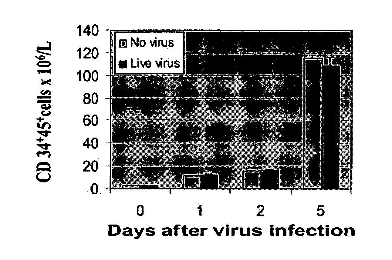
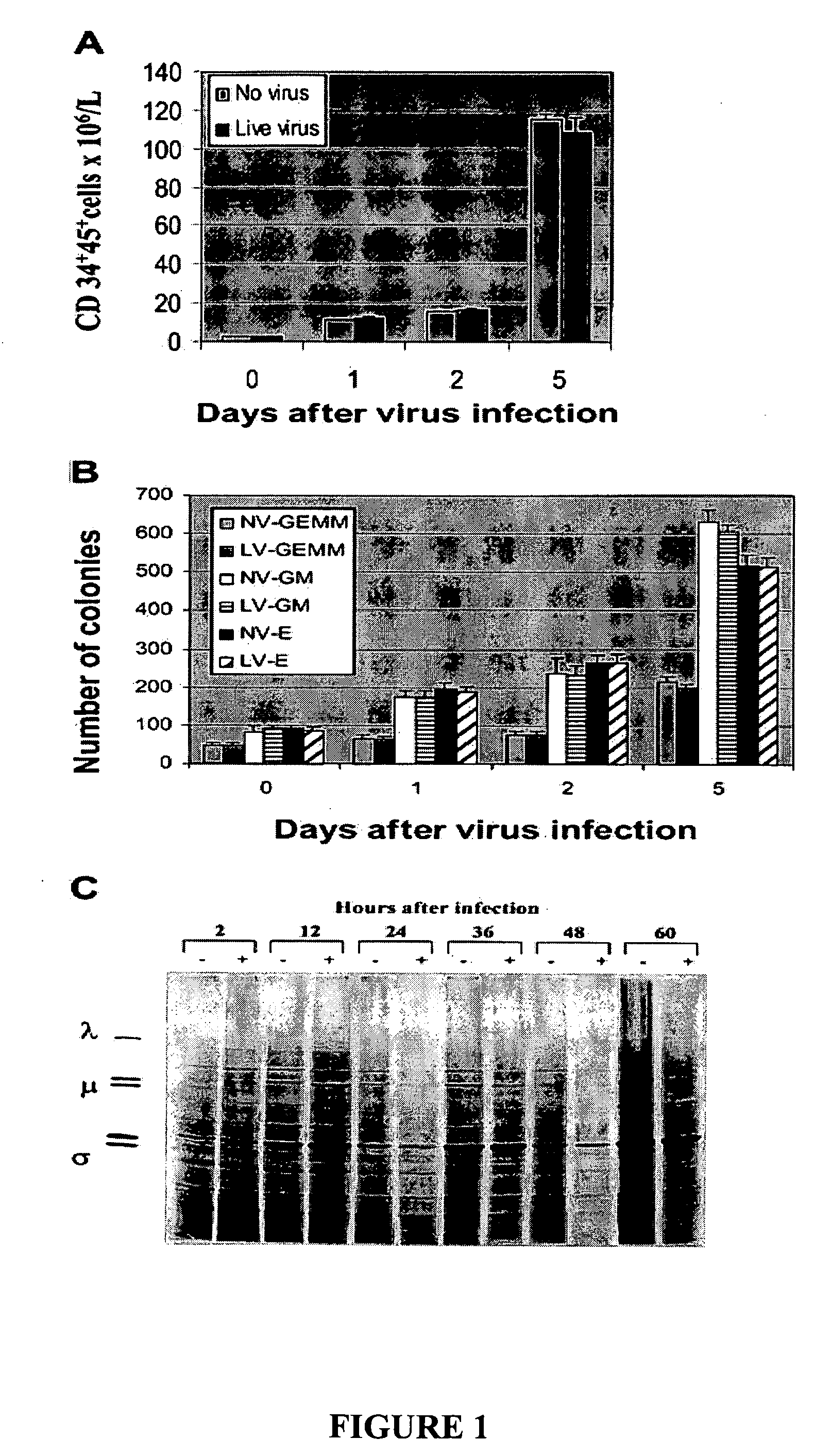
[0230] To investigate whether reovirus would affect the number and function of stem cells, positively selected (CD34+) stem cells were challenged with reovirus at an MOI of 40 and cultured in StemSpan medium for 5 days. As shown in FIG. 1A the number of CD34+CD45+ cells significantly increased with prolonged incubation and, at day 5, were 80-fold higher than at the start of the experiment. No significant difference between virus-treated and untreated stem cells was detected.

[0231] The preservation of the clonogenic potential of virus-treated and untreated hematopoietic progenitors was determined by culturing CD34+-enriched stem cells in StemSpan medium plated in methylcellulose medium. CFU-GMs, BFU-Es, and CFU-GEMMs were counted after 14 days of incubation. As shown in FIG. 1B, no differences in the clonogenic capacity of the stem cells was detected in virus-treated or untreated stem cells.

[0232] To confirm that reovirus does not ...

example 2

Flow Cytometric Analysis and [35S]-Methionine Labeling of Malignant Cell Lines

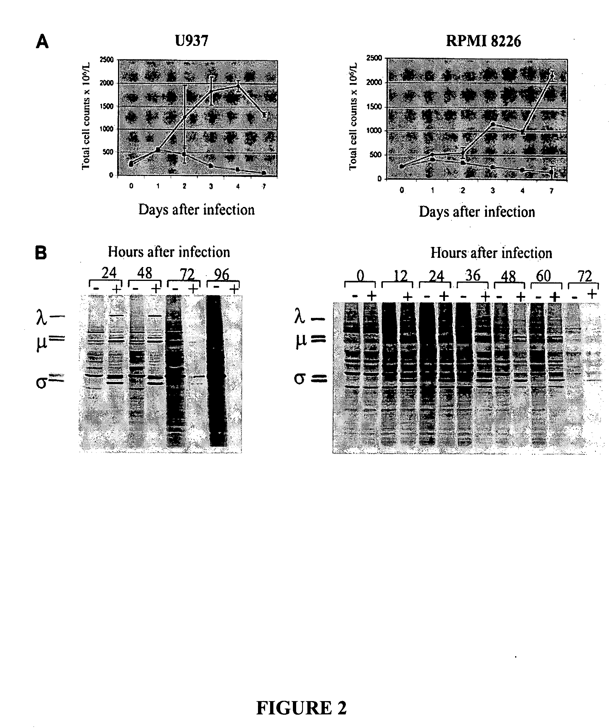
[0233] The susceptibility of established monocytic and myeloma cell lines to reovirus infection was tested by culturing U937 and RPMI 8226 cells in the presence (40 MOI) or absence of reovirus.

[0234] To confirm the cytopathic effect of reovirus on these cell lines samples of the cultured cells were obtained at days 0, 1, 2, 3, 4, and 7 days after virus infection and were analyzed for intact cell counts using propidium iodide. As shown in FIG. 2A, cell numbers declined after virus infection, contrasting the increase in uninfected cells. These results were confirmed over multiple experiments, and the cell counts approached zero by day 7. Residual cells seen at day 7 in FIG. 2A (left and right panels) are due to the fact that flow cytometry still counts membrane-intact but dying cells.

[0235] Replication of reovirus in susceptible cell lines was further confirmed by metabolic labeling with [35S]-methionine ...

example 3

Purging of Monocytic and Myeloma Cancer Cells in Apheresis Product

[0236] The results in Example 1 prove that exposure of hematopoietic stem cells to reovirus does not affect CD34+CD45+ cell counts or colony-forming potential of the hematopoietic progenitor cells in vitro. The monocytic and myeloma cancer cells were then mixed with apheresis product cells to result in tumor burdens of 1%, 0.1%, and 0.01% and purged with reovirus for 3 days. The purging efficacy of reovirus was evaluated using two different techniques: flow cytometry and cancer cell outgrowth following purging.

[0237] As depicted in FIG. 3Ai and 3Bi, reovirus treatment and purging for 3 days resulted in significant purging of U937 cells and complete purging at 0.01% contamination. When purged and unpurged admixed samples were recultured in RPMI medium for 6 days, no tumor regrowth was detected in the 0.1% and 0.01% contaminated samples (FIG. 3Ci). In contrast, U937 cell outgrowth was detected in all reovirus untreate...


PUM

PropertyMeasurementUnit
densityaaaaaaaaaa
cellular compositionaaaaaaaaaa
refractoryaaaaaaaaaa

Abstract

Oncolytic viruses can be used to purge cellular compositions to remove undesired neoplastic cells before the cellular compositions are used for transplantation. The present invention relates to the use of a virus to pre-treat a subject prior to delivery into the subject a transplant that has been purged with the same virus. This pre-treatment serves to elicit an immune response in the subject against the virus, thereby protecting the subject from infections by the virus after receiving the transplant, which likely contains infectious viruses.

Description

RELATED APPLICATION [0001] This application claims the benefit of U.S. Provisional Application No. 60 / 552,650, filed Mar. 12, 2004, which is herein incorporated by reference.TECHNICAL FIELD [0002] This invention relates to pre-treatment with a virus in combination with transplantation. REFERENCES [0003] U.S. Pat. No. 6,136,307. [0004] U.S. Patent Application Publication No. 20010048919. [0005] U.S. Patent Application Publication No. 20020006398. [0006] WO 94 / 18992, published Sep. 1, 1994. [0007] WO 94 / 25627, published Nov. 10, 1994. [0008] WO 99 / 08692, published Feb. 25, 1999. [0009] WO 99 / 18799, published Apr. 6, 2000. [0010] Alain, T., et al. (2002) Reovirus therapy of lymphoid malignancies. Blood. 100(12):4146-4153. [0011] Armitage, J. O. (1989). Bone marrow transplantation in the treatment of patients with lymphoma. Blood 73(7):1749-1758. [0012] Ball, E. D., et al. (1990). Autologous bone marrow transplantation for acute myeloid leukemia using monoclonal antibody-purged bone mar...

Claims


Application Information

Patent Timeline
Patent Type & Authority Applications(United States)
IPC IPC(8): A61K35/14A61K35/23A61K35/34A61K35/36A61K35/39A61K35/407A61K35/42A61K35/44A61K35/76A61K39/00A61K45/06A61K48/00C12N15/86
CPCA61K35/15A61K35/28C12N2720/12032A61K39/00A61K45/06A61K35/765
Inventor MORRIS, DONALDTHOMPSON, BRADLEY G.COFFEY, MATTHEW C.
Owner ONCOLYTICS BIOTECH
Why Eureka
Try Eureka